5137
Deformable registration of calf muscle MRI using an improved Demons approach1Electrical and Computer Engineering, University of Utah, Salt Lake City, UT, United States, 2Department of Radiology and Imaging Sciences, University of Utah, Salt Lake City, UT, United States, 3Department of Internal Medicine, Division of Geriatrics, University of Utah, Salt Lake City, UT, United States, 4Department of Internal Medicine, Division of Bioinformatics, University of Utah, Salt Lake City, UT, United States, 5Department of Internal Medicine, Division of Vascular surgery, University of Utah, Salt Lake City, UT, United States, 6Department of Internal Medicine, Division of Cardiology, University of Utah, Salt Lake City, UT, United States
Synopsis
Lower-extremity peripheral arterial disease (PAD) as a major clinical problem and MRI, which measures multiple aspects of the function of the calf muscles, such as muscle perfusion and oxygenation has not been significantly used for this. The reason being no efficient scanning and processing to compare data acquired during the course of treatment. Here we propose to register the calf MRI images using a modified Demons registration method which is more efficient. This method involves first a fast rigid registration and then the Demons deformable registration, by first applying rigid translation and rotation which substantially improved the registration performance for the calf muscle images.
Introduction
Lower-extremity peripheral arterial disease (PAD) as a major clinical problem affects 3 million patients per year in the United States. With PAD, peripheral blood vessels are narrowed so that blood flow to the affected limb is reduced. The ischemic state may lower the performance of the downstream calf muscles. MRI measures multiple aspects of the function of the calf muscles, such as muscle perfusion and oxygenation [4]. In applying these imaging techniques to the deformable calf, the calf is often imaged as different shapes in different scans, due to inconsistent positioning and coil set-up. For this reason, it is challenging to compare a same subject’s parameter maps acquired in different scans in a voxelwise manner.
In this abstract, we propose to register the calf MRI images using a modified Demons registration method. This method involves first a fast rigid registration and then the Demons deformable registration. For 8 human subjects, we implemented the registration method to register dynamic contrast enhanced (DCE) MRI data acquired on 2 different days.
Methods
Eight human subjects (6 male and 2 female, years 19-31) were recruited in this IRB-approved study. For each subject, MR scans were performed on a 3T MRI scanner (TrimTrio; Siemens Medical Solutions, Erlangen, Germany). A 4-channel flex coil was wrapped around the calf of one leg. With an in-scanner apparatus, the subject performed plantar flexion for 3 minutes at a frequency of 1 push per sec. A load of 4 lbs was attached to the exercise apparatus. Upon completion of the exercise, 0.05mmol/kg gadoteridol (Prohance; Bracco) was injected intravenously at a rate of 5ml/s. Then dynamic imaging started and continued for 4 minutes using a 2D saturation-recovery turboFLASH sequence: delay-time 300 ms, TR 527 ms, TE 1.42ms, flip angle 15°, slice thickness 10 mm, matrix 128×128, FOV 160×160 mm, temporal resolution 1s/frame. The MRI scan was repeated on a different day 1~3 weeks later.
Pre-registration by rigid translation and rotation: This pre-registration was to improve the performance and efficiency of the following Demons registration, which is more effective for registering local non-rigid deformations. In the rigid registration, the modified demons registration looks at 2 aspects of rigid transformations, translation and rotation. The translation is performed by comparing two centers which are obtained by computing the center of mass of the image. The rotation is performed by comparing the two centers taking 3 reference points and performing and affine transformation.
Non-rigid registration by the Demon’s method: Once two images were aligned to a same center and orientation, the Demons registration was applied to correct for local deformations. The Demons registration used optical flow to calculate the transformation model which maps the source image to target image. The optical flow is based on the gradient of the images, as proposed by Thirion[2]
$$ v=(m-s)*Δs / (|Δs^2| + (m-s)^2) $$ [1] where v is the displacement of the location of pixel and Δs is the gradient of the source image. Based on the optical flow, one image is transformed to match the other image in an iterative approach, and the iteration terminates when similarity between the two images is high enough.
Performance of the registration: To assess the similarity between the target image and the registered image, we computed the correlation between the two images. As comparison, the original Demons method (without preregistration) was also applied to the data, and the similarity metric was computed.
Results
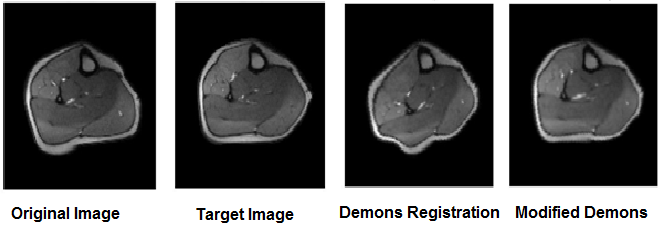
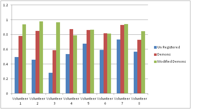
The proposed method was successfully applied to DCE MRI data of all 8 data sets. Figure 1 shows one representative example. The correlation coefficients of all the test images are shown in Figure 2. Compared to the correction coefficients 0.54±0.14 between the target and the unregistered images, both the original Demons and the modified Demons method provided significantly higher correlation, 0.80±0.11 and 0.89±0.07, respectively.Discussion
Our study suggests that the modified Demons registration, by first applying rigid translation and rotation, substantially improved the registration performance for the calf muscle images. In our implementation, there was one data set (volunteer 4 in Figure 2) where the correlation coefficient with the original Demons was higher than that of the modified Demons method. This was because the muscle groups activated differently between the two scans, and the intensity difference lowered the correlation coefficient for the modified method. In conclusion, the modified Demons registration is a promising method for efficiently registering calf muscle MR Images.Acknowledgements
No acknowledgement found.References
[1] Kroon, D-J., & Slump, C. H. (2009). MRI Modality transformation in demon registration. In IEEE International Symposium on Biomedical Imaging: From Nano to Macro, ISBI '09 (pp. 963-966). USA: IEEE Signal Processing Society. DOI: 10.1109/ISBI.2009.5193214
[2] J.P. Thirion, "Image matching as a diffusion process: an analogy with Maxwell’s demons," Medical Image Analysis, pp. 243-260, September 1998.
[3] Vivier PH, Storey P, Rusinek H, et al. Kidney function: glomerular filtration rate measurement with MR renography in patients with cirrhosis. Radiology. 2011;259:462–470.
[4] Zheng J, Hasting MK, Zhang X, et al. A pilot study of regional perfusion and oxygenation in calf muscles of individuals with diabetes with a non-invasive measure. J Vasc Surg. 2014;59:419–26.
Figures