5133
Diffusion, perfusion, and T2 analyses of lower leg muscles after intermittent pneumatic compression1Graduate School of Medical Sciences, Kanazawa University, Kanazawa, Japan, 2School of health Sciences, College of Medical, Pharmaceutical and Health Sciences, Kanazawa University, Kanazawa, Japan
Synopsis
Although the IPC supposedly changes regional muscle functions, this mechanism has not been clarified. Therefore, to quantitatively assess the effect of IPC, we simultaneously acquired functional information on diffusion, perfusion, and transverse relaxation time (T2) for lower-leg muscles before and after IPC using single-shot diffusion echo-planar imaging (SSD-EPI) with different b-values and echo times (TE). IPC reduces water molecule diffusion in the soleus muscle. Our method makes it possible to simultaneously obtain regional functional information on diffusion, perfusion, and T2 in lower-leg muscles before and after IPC.
INTRODUCTION
Intermittent pneumatic compression (IPC) of the lower leg muscles is used to increase venous outflow, reduce edema, and prevent venous thromboembolism.1 Although the IPC supposedly changes regional muscle functions, this mechanism has not been clarified. Therefore, to quantitatively assess the effect of IPC, we simultaneously acquired functional information on diffusion, perfusion, and transverse relaxation time (T2) for lower-leg muscles before and after IPC using single-shot diffusion echo-planar imaging (SSD-EPI) with different b-values and echo times (TE).MATERIALS AND METHODS
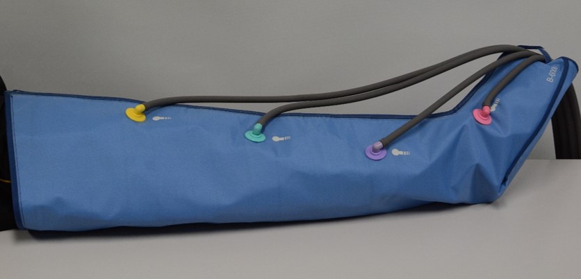
SSD-EPI with multiple b-values was performed on a 3.0-T MRI with the following imaging parameters: repetition time, 4000 ms; TE, 61.9 ms; field of view, 280 x 140 mm; imaging matrix, 128 x 128; slice thickness, 6 mm; number of signals averaged, 2; and b-values, 0, 25, 50, 200, 400, and 800 s/mm2. Under these conditions, we obtained transverse diffusion-weighted images at the maximum diameter of the right lower leg before and after IPC in five healthy volunteers. SSD-EPI with a b-value of 0 s/mm2 was also performed with a TE of 35.9 ms for T2 measurement. The experiment was initiated after the subject had rested in the supine position for 45 min to reduce the influence of postural changes on blood flow. Next, the treatment of the lower leg muscles were performed for 10 min with an IPC device (Fig. 1), and the same imaging was repeated immediately after IPC treatment. Then, we calculated perfusion-related and restricted diffusion coefficients (D* and D) with a two-step biexponential fitting2 for the gastrocnemius, tibialis anterior, and soleus muscles. T2 of each muscle was calculated from b0 images with different TEs.RESULTS AND DISCUSSION
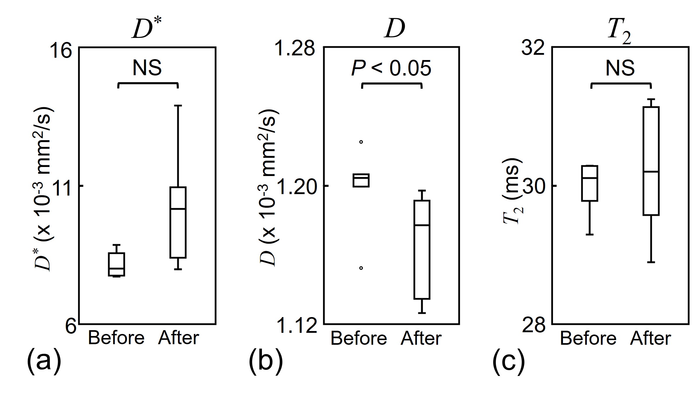
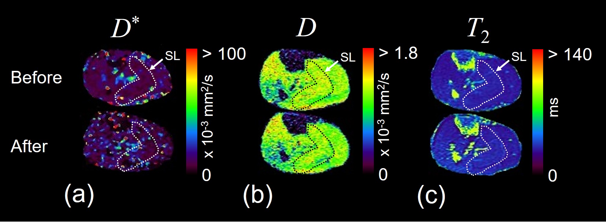
D of the soleus muscle was significantly lower than that before IPC (P < 0.05) (Figs. 2 and 3). However, there was no significant difference in D*, D, or T2 of the other lower leg muscles before and after IPC. These results indicate that the effect of IPC depends on the muscle composition, for instance, the soleus muscle contains many slow-muscle fibers.3CONCLUSION
IPC reduces water molecule diffusion in the soleus muscle. Our method makes it possible to simultaneously obtain regional functional information on diffusion, perfusion, and T2 in lower-leg muscles before and after IPC.Acknowledgements
No acknowledgement found.References
1. Chen AH, et al., Eur J Vasc Endovasc Surg. 2001; 21: 383-392.
2. Ohno N, et al., J Magn Reson Imaging. 2016; 43: 818-823.
3. Moore KL, et al., Clinially Oriented ANATOMY FIFTH EDITION. 2006; 648.
Figures