5115
Assessing changes in kidney pH in acute kidney injury model using acidoCEST MRI1School of Engineering, Macquarie University, Sydney, Australia, 2Centre for Pre-Clinical Imaging, University of Liverpool, Liverpool, United Kingdom, 3Department of Medical Imaging, University of Arizona, Tucson, AZ, United States, 4Department of Cancer System Imaging, MD Anderson Cancer Centre, Houston, TX, United States
Synopsis
Kidneys are responsible for regulation of pH homeostasis, and cytotoxicity caused by cancer therapeutics can significantly alter renal function and homeostasis. Chemical exchange saturation transfer (acidoCEST) MRI has been proposed to measure tissue pH in-vivo using exogenous contrast agents. In this study, we used the acidoCEST technique to measure changes in kidney pH after acute kidney injury (AKI) in rodents. Typically, CT contrast agents such as Iopamidol (300 mg iodine/mL) are used as CEST contrast agent in acidoCEST MRI. However, the accuracy of acidoCEST using CT contrast agents relies on the delivery of the contrast agent to the target organ. To address this issue, we performed acidoCEST and FAIR-EPI based perfusion imaging to assess pH and blood flow changes in a mouse model of AKI. Results show that perfusion of kidneys affect pH measurements.
Introduction
Kidneys regulate pH homeostasis, and cytotoxicity caused by cancer therapeutics can significantly alter renal function and homeostasis. We have used pH sensitive chemical exchange saturation transfer (acidoCEST) MRI methods to measure changes in kidney pH after acute kidney injury (AKI) in rodents (1-2). However, the accuracy of acidoCEST using CT contrast agents relies on the delivery of the contrast agent to the target organ. To address this issue, we performed acidoCEST and FAIR-EPI based perfusion imaging to assess pH and blood flow changes in a mouse model of AKI.Methods
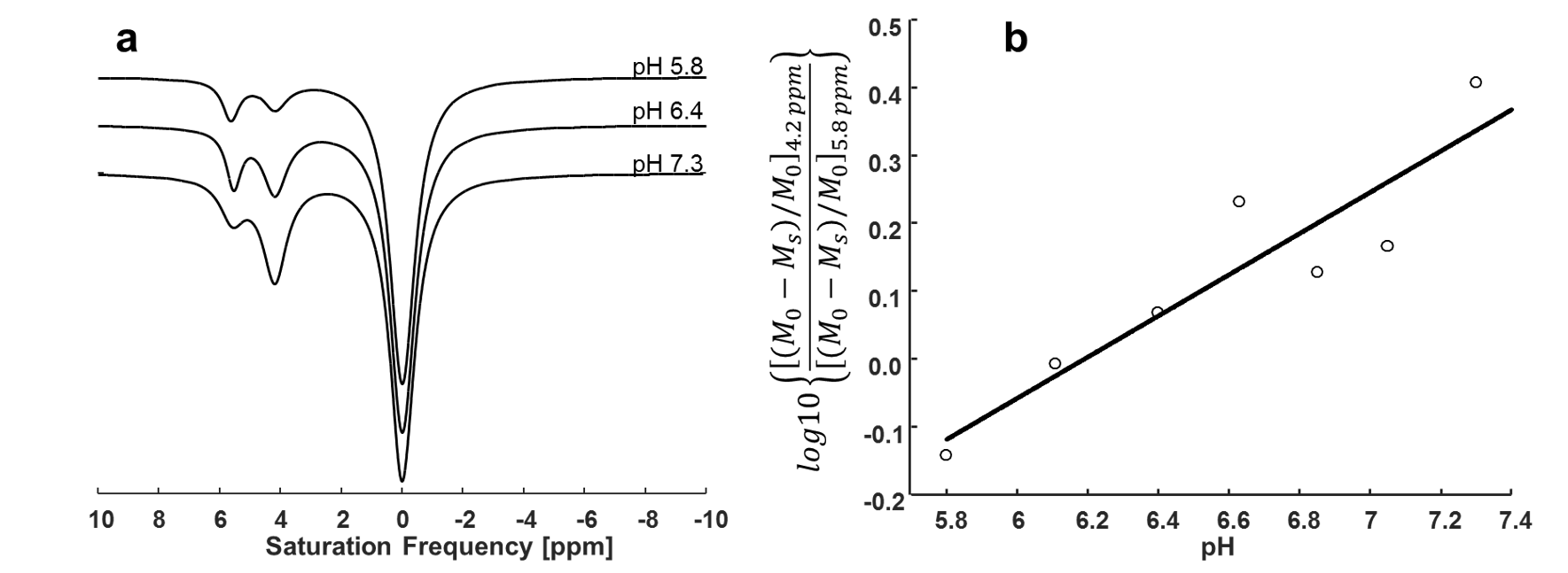
AKI was induced in 7 male BALB/c mice (6–8-week old), by i.p. injection of adriamycin (20 mg/kg body weight). Saline was injected in 7 control animals. Iopamidol (300 mg iodine/mL) was used as CEST contrast agent. All MRI experiments were carried out using a 9.4 Tesla Bruker scanner with a 27-mm diameter quadrature volume coil (PulseTeq). A CEST-FISP MRI sequence was used for data acquisition with: TR/TE = 3.26/1.63 ms; flip angle = 60; in-plane resolution = 234µm2; slice thickness = 2mm; field of view = 30×30mm2; linear encoding order; unbalanced FID mode; and 418.26 ms scan time. A series of 60 MR frequencies were saturated to acquire a CEST spectrum without and with iopamidol injection. The pre-injection data was subtracted from post-injection data to remove the CEST effects from endogenous agents. This difference was fitted with Lorentzian line shapes centered at 4.2 and 5.8 ppm. A logarithmic ratio of CEST peaks at these two frequencies was obtained (2). This ratio was compared with the pH calibration curve obtained using similar data processing steps for the samples prepared using iopamidol. The in vivo pH of both kidneys was calculated using this calibration curve. All in vivo experiments were performed at 34.5 ± 1.50C. FAIR-EPI was performed with same location and resolution as CEST-FISP and TR/TE = 12000/16.82 ms; inversion time = [26, 400, 800, 1200, 1600, 2000, 2400, 2800, 3200, 3600, 4000, 4400, 4800, 5200, 5600, 6000] ms.Results
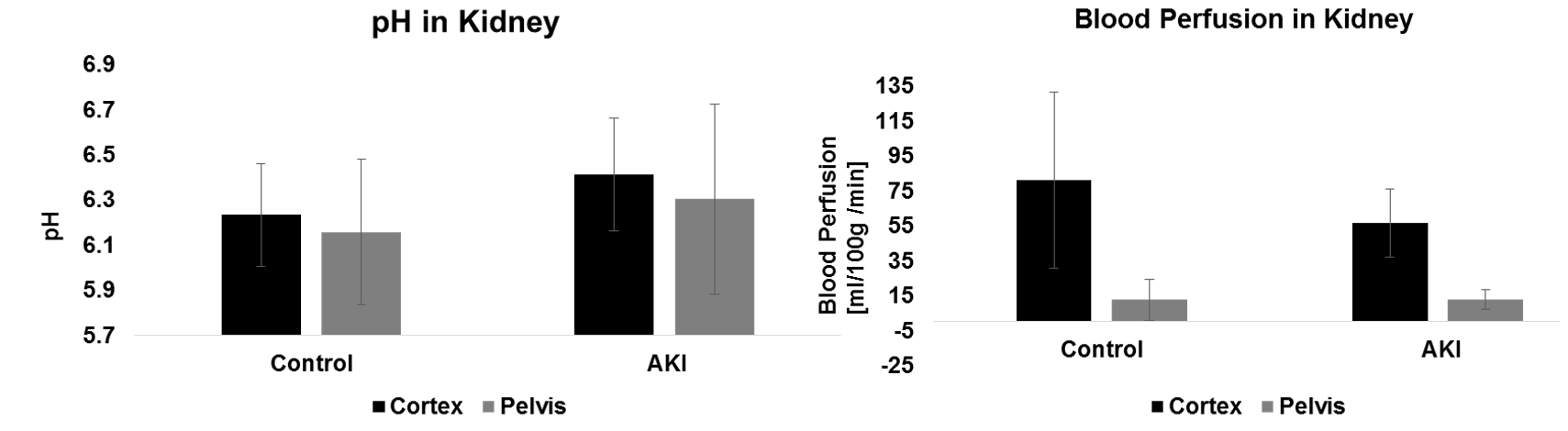
The pH sensitivity of the CEST effect is shown in Figure 1a. A linear pH calibration curve was obtained from iopamidol solutions as shown in Figure 1b. Our analysis revealed a non-significant increase in pH in the cortex and pelvis of kidneys four days after adriamycin injection when compared to controls (p=0.37 for cortex, and p=0.38 for pelvis) (Figure 2a). FAIR perfusion results for the same animals showed a significant reduction in blood flow in the cortex (p=0.05) but no change in the renal pelvis (p=0.18) (Figure 2b).Discussion and Conclusion
Here, we have explored the use of acidoCEST and FAIR-EPI measurements to determine pH and blood flow changes in kidneys of mice with adriamycin-induced AKI. Previously, a maximal increase in kidney pH had been reported on day three in a glycerol induced AKI model (1). The insignificant pH increase on day four in our studies could be based on different levels of kidney injury due to adriamycin in comparison to glycerol. The higher variation of pH values in pelvis could be attributed to reduced blood flow which led to a decrease in the concentration of iopamidol in the pelvis thereby reducing the signal-to-noise ratio in pH measurements. These results show that in areas of higher blood flow such as cortex, the acidoCEST measurements are reliable, but maybe questionable in poorly perfused regions such as pelvis.Acknowledgements
The work was funded by Early Career Researchers and Returners Fund, University of Liverpool, United Kingdom.References
1. D L Longo et al. Magn Reson Med 2013; 70: 859-864.
2. B F Moon et al. Contrast Media Mol Imaging 2015; 10: 446-455
Figures