5083
In vivo Current Density and Conductivity Tensor Imaging of Human Brain During TACS using DT-MREIT1SBHSE, Arizona State University, Tempe, AZ, United States, 2Department of Clinical and Health Psychology, University of Florida, Gainesville, FL, United States, 3GE Healthcare, Wausau, WI, United States, 4Dept. of Biochemistry & Molecular Biology, University of Florida, Gainesville, FL, United States
Synopsis
Knowledge of the electrical properties of brain tissue is key to developing better understanding of whole brain function. In this study, we present the first in vivo images of anisotropic conductivity distribution in the human head, measured at a frequency of ~10 Hz. We used MREIT techniques to encode phase changes caused by transcranial AC current flow (TACS) within the head via two independent electrode pairs. These results were then combined with DTI data to reconstruct full anisotropic conductivity distributions in 5 mm-thick slices of the brains of two participants. Conductivity values recovered in the study were broadly consistent with literature values.
Introduction
The electrical conductivity distribution inside the human head controls pathways of external or internal currents. Accurate measurement of these conductivities is key to developing better understanding of whole brain function. In neuromodulation techniques such as transcranial DC or AC stimulation, access to accurate conductivity distributions may improve targeting of different cortical structures. While in-vivo reports of conductivity measurements in human heads are limited, low-frequency MR electrical impedance tomography (MREIT)1 methods make it possible to recover current density distributions in subjects using one component (Bz) of magnetic flux density vectors. A recent extension of MREIT methods, diffusion tensor-MREIT (DT-MREIT)2 can be used to reconstruct full anisotropic conductivities and current density distributions using MREIT and diffusion tensor image data gathered from the same subject, and has recently been demonstrated in canines3. In this study, we present the first conductivity reconstruction images from a human subject obtained from in-vivo DT-MREIT imaging during TACS.Methods
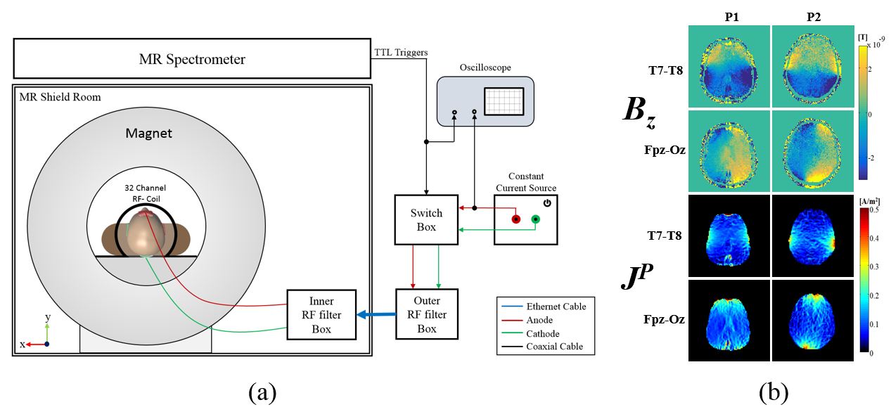
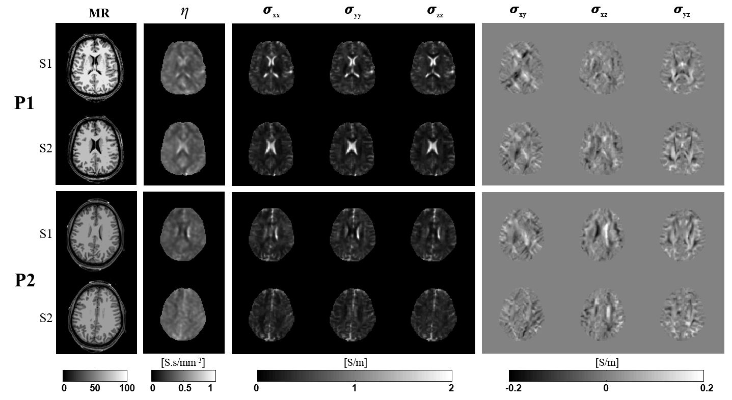
All procedures were performed according to protocols approved by the University of Florida (UF) and Arizona State University Institutional Review Boards. Two healthy participants were imaged in 3T MRI scanner during TACS. A current intensity of 1.5mA with a frequency of ~10 Hz was applied to the subject’s head via surface electrodes (~36cm2) using T7-T8 and FPz-Oz montages. T1-weighted, MREIT and DWI data were obtained in the same imaging session, following a previously published protocol4 and co-registered for image reconstruction. DWI data was processed to diffusion tensors using FSL. Projected current densities JP were recovered from MREIT Bz using techniques described previously5 then used to calculate a diffusivity ratio2, η. Finally, conductivity tensors were obtained by multiplying diffusion tensors by η distributions.Results and Discussions
Figure 1a shows a schematic of the measurement setup. Experimental measured Bz, and computed JP are shown in Figure 1b. Figure 2 shows reconstructed MR magnitude, η and conductivity tensor images of two slices of each participant brain using DT-MREIT methods. Calculated reconstructed conductivity values in ROIs selected within CSF and gray matter were 1.583 S/m and 0.287 S/m respectively. White matter conductivity values were 0.391 S/m and 0.132 S/m for longitudinal and transverse directions, respectively, and 0.218 S/m on average.
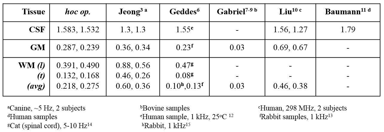
These conductivity values were consistent with reported conductivity values form measurements performed in human and animal samples. We compare the values measured here with those reported in other contexts and studies. A summary of this survey is presented in Figure 3 below. In Figure 3, tissue conductivities cited were measured in vivo, at body temperature and at 10 Hz unless otherwise specified.
Conclusion
We reconstructed conductivity tensors from in vivo DT-MREIT imaging during TACS in the human head. DT-MREIT conductivity values were consistent with values found in the literature. Applications of our methods and findings will assist model validation in transcranial electrical stimulation and EEG source localization problems. Future studies will involve pulse sequence acceleration to maximize brain coverage and resolution.Acknowledgements
Research reported in this abstract was supported by NIH awards R21NS081646 and RF1MH114290 to RJS. A portion of this work was performed in the McKnight Brain Institute at the National High Magnetic Field Laboratory’s AMRIS Facility, which is supported by National Science Foundation Cooperative Agreement No. DMR-1157490 and the State of Florida.References
[1] Woo EJ, Seo JK. Magnetic resonance electrical impedance tomography (MREIT) for high-resolution conductivity imaging. Physiol Meas 2008;29; R1–R26.
[2] Kwon OI, Sajib SZK, Sersa I, et al. Current density imaging during transcranial direct current stimulation (tDCS) using DT-MRI and MREIT: Algorithm development and numerical simulations. IEEE Trans Biomed Eng 2015;63(1);167-75.
[3] Jeong WC, Sajib SZK, Katoch N, et al. Anisotropic conductivity tensor imaging of canine brain using DT-MREIT. IEEE Trans Med Imaging 2017;36(1);124-131.
[4] Kasinadhuni AK, Indahlastari A, Chauhan M, et al. Imaging of current flow in the human head during transcranial electrical therapy. Brain Stimul 2017;10(4):764-772.
[5] Park C, Lee BI, Kwon OI. Analysis of recoverable current from one component of magnetic flux density in MREIT and MRCDI. Phys Med Biol 2007;52; 3001–3013.
[6] Geddes LA, Baker LE. The specific resistance of biological materials: a compendium of data for the biomedical engineer and physiologist. Med Biol Eng 1967;5(3):271-93.
[7] Gabriel C1, Gabriel S, Corthout E. The dielectric properties of biological tissues: I. Literature survey. Phys Med Biol 1996;41(11):2231-49.
[8] Gabriel S, Lau RW, Gabriel C. The dielectric properties of biological tissues: II. Measurements in the frequency range 10 Hz to 20 GHz. Phys Med Biol 1996;41(11):2251-69.
[9] Gabriel S, Lau RW, Gabriel C. The dielectric properties of biological tissues: III. Parametric models for the dielectric spectrum of tissues. Phys Med Biol. 1996 Nov;41(11):2271-93.
[10] Liu J, Zhang X, Schmitter S, et al. Gradient-based electrical properties tomography gEPT): a robust method for mapping electrical properties of biological tissues in vivo using magnetic resonance imaging. Magn Reson Med 2015;74(3):634-46.
[11] Baumann SB, Wozny DR, Kelly SK, et al. The electrical conductivity of human cerebrospinal fluid at body temperature. IEEE Trans Biomed Eng 1997;44(3):220-3.
[12] Radvan-Ziemnowicz SA, McWilliams JC, Kucharski WE. Conductivity versus frequency in human and feline cerebrospinal fluid. Proc. 17th Ann Conf Eng Med Biol 1964; 6;108.
[13] Crile GW, Hosmer HR, Rowland AF. The electrical conductivity of animal tissues under normal and pathological conditions. American Jour of Physiology 1922; 60; 59–106.
[14] Ranck JB, BeMent SL. The specific impedance of the dorsal columns of cat: an anisotropic medium. Experimental Neurology 1965;11; 451–463.
[15] Harreveld A, Murphy T, Nobel KW. Specific impedance of rabbit’s cortical tissue,” American Journal of Physiology 1963; 205; 203–207.
Figures