5069
Rapid Measurement of ATP Kinetics in Human Skeletal Muscle at 7T1Advanced Imaging Research Center, University of Texas Southwestern Medical Center, Dallas, TX, United States, 2Department of Radiology, University of Texas Southwestern Medical Center, Dallas, TX, United States, 3Department of Chemistry, University of Texas at Dallas, Richardson, TX, United States, 4VA North Texas Health Care System, Dallas, TX, United States
Synopsis
Measuring ATP energy metabolism in human subjects by conventional 31P saturation transfer (ST) is challenging because of multiple competing metabolic pathways, high SAR, and artifacts caused by the prolonged B1-saturation pulses. EKIT (exchange kinetics by inversion transfer) allows magnetization transfer (MT) to evolve in the absence of B1 pulsing and multiple pathways can be probed by inverting all spins individually, but the process is time consuming and MT effects are small. A multi-module EBIT technique (exchange kinetics by band inversion transfer) addresses these issues and provides a comprehensive picture of ATP kinetics in human skeletal muscle.
INTRODUCTION
An adequate ATP supply is essential for normal muscle function. Reduced myocellular ATP production has been associated with mitochondrial dysfunction and insulin resistance, a strong risk factor for developing type 2 diabetes.1,2 A widely used non-invasive technique to measure ATP synthesis is 31P saturation transfer (ST).3 However, because of co-existence of multiple competing kinetics pathways, the typically long B1-saturation duration required for achieving steady state MT effect is associated with high SAR and prone to artifacts from off-resonance saturation and activation of small hidden pools of phosphates.4 To overcome these issues, inversion transfer (IT) methods, which enable MT effect to buildup in the absence of B1 pulsing, have been proposed, 5-7 including exchange kinetics by inversion transfer (EKIT), by which all individual spins are selectively inverted to activate connected MT pathways in the exchange system. But EKIT is time-consuming and the MT effect is typically small. To address this issue, we took advantage of existing EBIT (exchange kinetics by band inversion transfer) method,6,7 which is capable of enhancing MT effect in a selected pathway by inverting a group of spins, and developed a short-TR multi-module EBIT approach, termed mEBIT, for rapid measurement and ATP kinetics in multiple pathways, as demonstrated here in human skeletal muscle at 7T.
METHODS
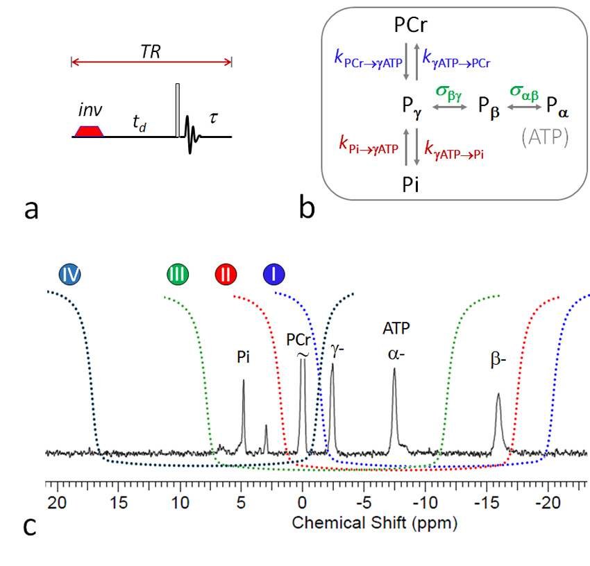
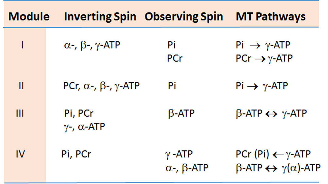
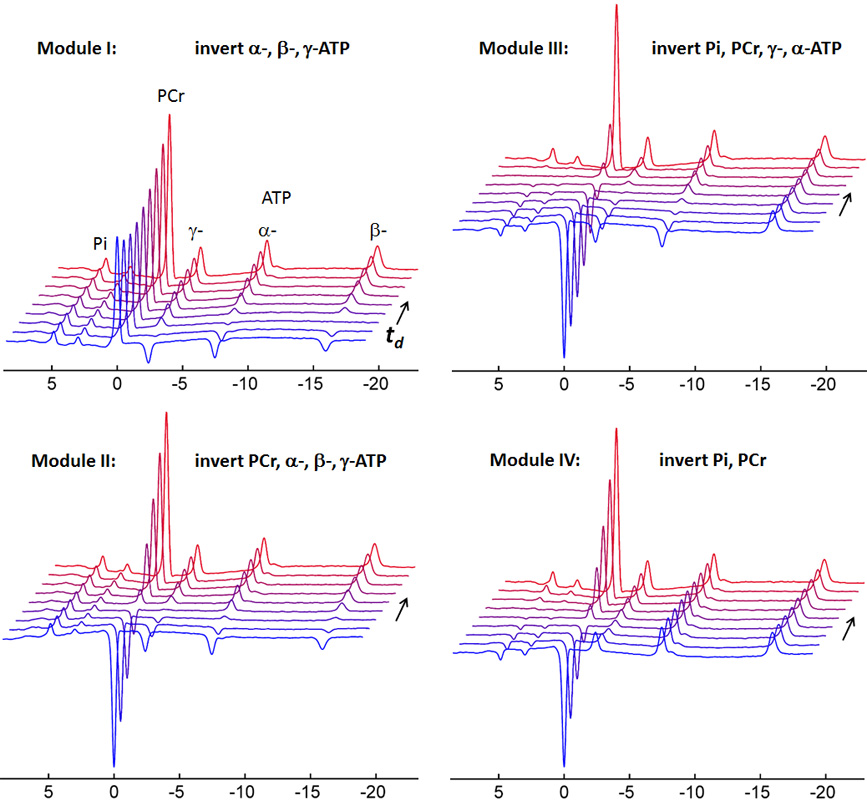
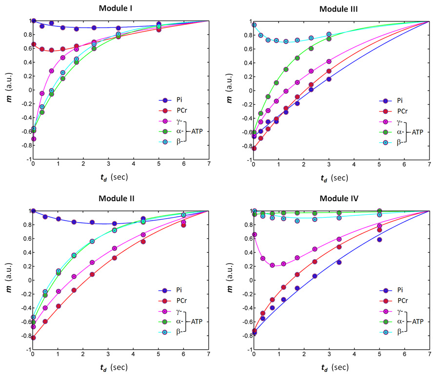
Five healthy subjects participated in the study with written consent under an IRB-approved protocol. The 31P spectra were acquired from calf muscle at 7T (Philips Healthcare) using a 1H/31P T/R coil (φ=10 cm) with four mEBIT modules, denoted as Modules I, II III and IV. These four mEBIT modules differ in center frequency of the inversion pulse which is specifically designed for inverting selected groups of 31P spins (Figures 1 and 2). A basic EBIT sequence was composed of an adiabatic inversion pulse followed by a delay period td , in range 0.025 – 6.0 sec (8 data points), and a hard readout pulse while keeping TR constant (7 sec). Scan time for each mEBIT module was 7.5 min with NA = 8. One single reference scan was also collected without inversion pulse under otherwise the same condition as mEBIT. The Z-magnetization of each 31P signal (Mz) measured by mEBIT was normalized by the Z-magnetization (Mz*) obtained from the reference scan (Mz*) with m = Mz/Mz*. All normalized m-data were fitted to a five-spin model including Pi, PCr, α-, β- and γ-ATP using mCal, an in-house developed Matlab program, based on matrix equations as described in ref [8].RESULTS and DISCUSSION
Figure 3 shows a representative 31P mEBIT dataset acquired from calf muscle of a healthy subject. The m ~ td data for the exchange system fit well to the 5-spin exchange model as shown by the curve fitting (Figure 4). Using these mEBIT modules, a comprehensive picture of ATP kinetics was obtained, with large MT effects observed at Pi and PCr for forward ATP synthesis reactions Pi→γ-ATP (Modules I and II) and PCr → γ-ATP (Module I), and at γ-ATP by reverse reactions γ-ATP→Pi and γ-ATP→ PCr (Module IV, Figures 3 and 4). In addition, ATP intramolecular 31P-31P NOE effect was also clearly identifiable at β-ATP both as direct MT effect through γ-ATP and α-ATP pathways (Module III), and as relayed MT effect through PCr (Pi)→γ-ATP→β-ATP pathway (Module IV, Figure 2 and 3). As compared to EKIT,5 mEBIT dramatically improved both MT effect (typically 2 – 3 fold) and SNR (one order of magnitude under current condition). For the group of subjects studied (N =5), the following kinetic rate constants and relaxation times were obtained for resting calf muscle: de novo synthesis kPi→γATP = 0.063 ± 0.013 sec-1, CK-mediated ATP synthesis kPCr→γATP = 0.31 ± 0.06 sec-1, ATP cross-relaxation (NOE effect): σγ(α)↔βATP = -0.19 ± 0.03 sec-1, T1(Pi) = 9.4 ± 1.4 sec, T1(PCr) = 6.2 ± 0.4 sec, T1(γ-ATP) = 1.9 ± 0.1 sec, T1(α-ATP) = 1.4 ± 0.1 sec and T1(β-ATP) = 1.1 ± 0.1 sec, in agreement with results by EKIT,5 and by long-TR (30 sec)single-module EBIT for de novo ATP synthesis,7 but with significantly reduced scan time (~ 30 min), as compared to EKIT (~ 1 hr).5 Furthermore, when available scanner time is constrained, mEBIT offers scan flexibility, by eliminating modules with redundant MT pathways (Figure 2). For example, a combination of Module I and IV (2-module mEBIT) can cut scan time to 15 min. With a trade-off in SNR by a factor of 1.4 (reducing NA from 8 to 4), one can further cut scan time to 7.5 min.CONCLUSIONS
mEBIT allows fast and comprehensive evaluation of ATP exchange kinetics in skeletal muscle, and it could become a useful tool for measuring energy metabolism in patients on clinical 7T scanners.Acknowledgements
No acknowledgement found.References
1. Petersen KF, Dufour S, Shulman GI. Decreased insulin-stimulated ATP synthesis and phosphate transport in muscle of insulin-resistant offspring of type 2 diabetic parents. PLoS Med. 2005;2(9):e233.
2. Petersen KF, Dufour S, Befroy D, Garcia R, Shulman GI. Impaired mitochondrial activity in the insulin-resistant offspring of patients with type 2 diabetes. N Engl J Med. 2004;350(7):664-71.
3. Lei H, Ugurbil K, Chen W. Measurement of unidirectional Pi to ATP flux in human visual cortex at 7 T by using in vivo 31P magnetic resonance spectroscopy. Proc Natl Acad Sci U S A. 2003;100(24):14409-14.
4. Balaban RS, Koretsky AP. Interpretation of 31P NMR saturation transfer experiments: what you can't see might confuse you. Focus on “Standard magnetic resonance-based measurements of the Pi→ATP rate do not index the rate of oxidative phosphorylation in cardiac and skeletal muscles”. Am J Physiol Cell Physiol 2011;301:C12–C15.
5. Ren J, Yang B, Sherry AD, Malloy CR. Exchange kinetics by inversion transfer: integrated analysis of the phosphorus metabolite kinetic exchanges in resting human skeletal muscle at 7 T. Magn Reson Med. 2015;73(4):1359-69.
6. Ren J, Sherry AD, Malloy CR. 31P-MRS of healthy human brain: ATP synthesis, metabolite concentrations, pH, and T1 relaxation times. NMR Biomed. 2015;28(11):1455-62.
7. Ren J, Sherry AD, Malloy CR. Amplification of the effects of magnetization exchange by (31) P band inversion for measuring adenosine triphosphate synthesis rates in human skeletal muscle. Magn Reson Med. 2015;74(6):1505-14.
8. Ren J, Sherry AD, Malloy CR. Efficient 31 P band inversion transfer approach for measuring creatine kinase activity, ATP synthesis, and molecular dynamics in the human brain at 7 T. Magn Reson Med. 2017;78(5):1657-1666.
Figures