5062
Reproducibility of Diffusion Tensor Imaging (DTI) in the hamstrings of healthy athletes1Radiology & Nuclear Medicine, Academic Medical Center, Amsterdam, Netherlands, 2University Medical Center Utrecht, Utrecht, Netherlands
Synopsis
Muscle injuries are diagnosed using T2-weighted scans, but these techniques lack specificity for assessing tissue repair. DTI seems more suitable for this purpose, but reproducibility data is lacking. Therefore, the aim of this study was to determine the reproducibility of DTI, expressed as the within subject CV per DTI parameter in the hamstrings of healthy athletes. The wsCV values reported here for DTI parameters are superior or similar to previously reported wsCV. In conclusion, our protocol allows us to perform DTI on both upper legs simultaneously with an overall high SNR and high reproducibility.
Introduction
Muscle injuries comprise a large percentage of sports related injuries and are conventionally assessed using T2-weighted MR scans. However, current T2-weighted imaging techniques lack specificity for assessing tissue repair and predicting return to play1,2. Diffusion Tensor imaging (DTI) can probe muscle fiber micro-anatomy and has shown to be sensitive to muscle changes that remained undetected on conventional T2-weighted MRI3. Therefore DTI is suggested as an advanced diagnostic tool for assessing hamstring injuries. If both upper legs are scanned simultaneously in muscle injury studies, the muscles in the uninjured leg can function as a control. However, reaching sufficient data-quality in both legs using a bilateral protocol is challenging due to B0 and B1+ field inhomogeneities4. As a result, most DTI studies in the upper legs obtain data from only one leg. In addition, reference values for the reproducibility of DTI parameter measurements in the hamstrings of healthy athletes are lacking. Therefore, the aim of this study was to determine the reproducibility of DTI parameters in the hamstrings of both legs in healthy athletes over a period of 2 weeks.Methods
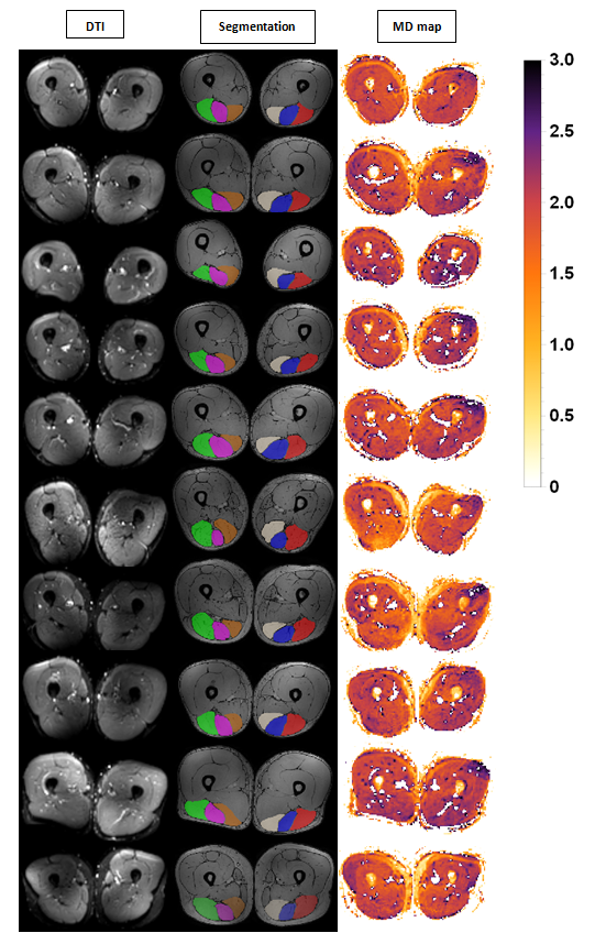
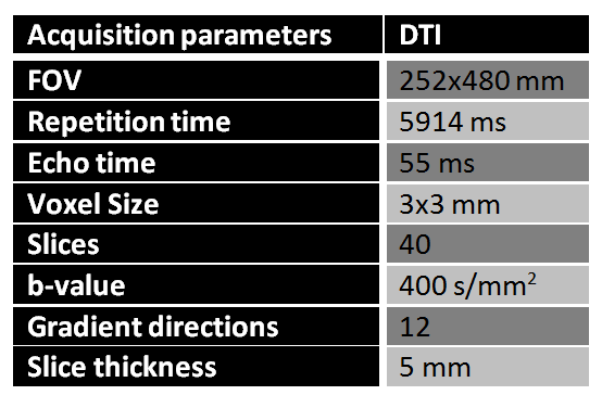
This study was approved by the local IRB and written informed consent was provided. DTI datasets (See Fig. 1) were acquired in the upper legs of 10 healthy athletes (9 males-1 female, age range: 22-40, mean age: 28.2, sports activity: 3x/week). Scans were acquired at 2 time points, 2 weeks apart (day 1 and day 14). A 3T MRI system was used (Ingenia; Philips Healthcare, Best, the Netherlands). Acquisition parameters are listed in Table 1. DTI datasets were processed using DTITools for Wolfram Mathematica (github.com/mfroeling/DTITools). The DTI data was denoised, registered and corrected for eddy currents and subject motion. A iterative weighted linear least squares (iWLLS) approach was used for the estimation of λ1, λ2 and λ3 from which Mean diffusivity (MD) and fractional anisotropy (FA) were calculated. The SNR per muscle was calculated in order to assess DTI quality using a noise map and the b = 0 s/mm2 data. ROIs were manually drawn for the Biceps femoris, Semimembranosus and Semitendinosus muscles in the right and left legs. DTI parameters and SNR are reported per time point per hamstring muscle as the mean value over the 20 middle slices. Muscles with SNR<20 at b = 0 s/mm2 were excluded from the analysis. The statistical analyses comprised one sample T-tests, Bland-Altman plots and within subject coefficient of variation (wsCV).Results
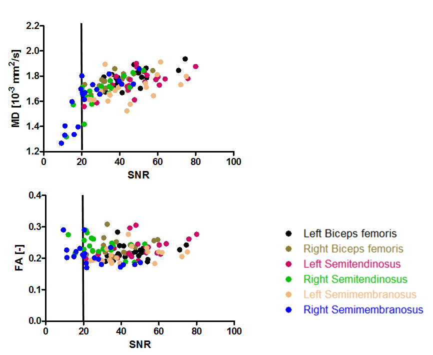
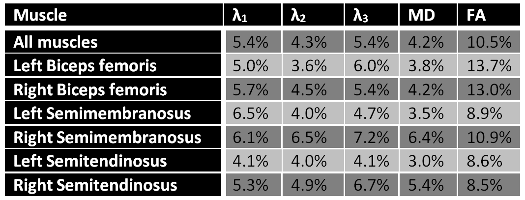
10 data points were excluded from analysis because the SNR was below 20. The Bland Altman plots showed good reproducibility for all DTI parameters (See Figure 2). The within subject Coefficient of Variation (wsCV) varied between 4.1% and 6.5% for λ1, 3.6% and 6.5% for λ2, 4.1% and 7.2% for λ3, 3.0% and 6.4% for MD and between 8.5% and 13.7% for FA (See Table 2). The wsCV values were similar for the left and right leg and no left-right bias was detected for λ1, λ2, λ3, MD, and FA. (one sample T-tests: MD: p=0.549, FA: p=0.012, λ1: p=0.450, λ2: p=0.404 and λ3: p=0.037). Figure 3 shows an underestimation of MD in the lower SNR range. There seems to be no overestimation of FA in the lower SNR range.Discussion
Our protocol allows us to perform DTI on both upper legs simultaneously with an overall high SNR (See Figures 1 and 3) and high reproducibility for all DTI parameters. This is demonstrated by the comparable range in wsCV values between the left and right leg. The range of reported wsCV values for FA match the values previously reported in the forearm at 3.0T and the human calf at 1.5T5,6. The wsCV values for λ1, λ2, λ3 and MD are superior to previously reported wsCV, which suggests a higher DTI quality compared to previous studies. There are no DTI reproducibility studies of the upper legs reported in literature for comparison.Conclusion
Taken together, these results show that our DTI protocol and post-processing is robust in both legs and therefore could be applied to assess whether small changes in DTI parameters due to muscle injury can be detected.Acknowledgements
This project was supported by the ZonMW Sportinnovator grant.References
1. Reurink, G., Whiteley, R. & Tol, J. L. Hamstring injuries and predicting return to play: ‘bye-bye MRI?’. Br. J. Sports Med. 49, 1162–3 (2015).
2. Horst, N., Hoef, S., Reurink, G., Huisstede, B. & Backx, F. Return to Play After Hamstring Injuries : A Qualitative Systematic Review of Definitions and Criteria. Sport. Med. (2016). doi:10.1007/s40279-015-0468-7
3. Froeling, M. et al. Muscle Changes Detected by Diffusion-Tensor Imaging after Long-Distance Running. Radiology 274, 140702 (2014).
4. Brink, W. M., Versluis, M. J., Peeters, J. M., Börnert, P. & Webb, A. G. Passive radiofrequency shimming in the thighs at 3 Tesla using high permittivity materials and body coil receive uniformity correction. Magn. Reson. Med. 76, 1951–1956 (2016).
5. Froeling, M. et al. Reproducibility of diffusion tensor imaging in human forearm muscles at 3.0 T in a clinical setting. Magn. Reson. Med. 64, 1182–1190 (2010).
6. Sinha, S. & Sinha, U. Reproducibility analysis of diffusion tensor indices and fiber architecture of human calf muscles in vivo at 1.5 Tesla in neutral and plantarflexed ankle positions at rest. J. Magn. Reson. Imaging 34, 107–119 (2011).
Figures