5057
Evaluation of Resting-State BOLD MRI of Calf Muscles in Healthy Volunteers and Patients with Peripheral Arterial Disease1Department of Radiology, Renji Hospital, School of Medicine, Shanghai Jiao Tong University, Shanghai, China, 2Department of Vascular Surgery, Renji Hospital, School of Medicine, Shanghai Jiao Tong University, Shanghai, China, 3GE Healthcare China, Shanghai, China
Synopsis
BOLD MRI is a helpful imaging modality for assessing tissue oxygenation/perfusion characteristics in skeletal muscles. However, few studies have explored the utility of resting-state BOLD MRI in healthy subjects and patients with peripheral arterial disease (PAD). We conducted this study aimed at addressing this question, and results showed that (1) T2* was found to be independent of age factor in calf muscles; (2) T2* was useful for differentiating PAD patients with age-matched older healthy subjects, as well as mild-to-moderate PAD patients with severe ones; and (3) T2* correlated well with ankle-brachial index in PAD patients.
Purpose
BOLD MRI of the skeletal muscle has shown potential in assessment of tissue oxygenation/perfusion in healthy volunteers and patients with peripheral arterial disease (PAD).1 Several dynamic paradigms have been employed to study the BOLD signal changes under different stimulation conditions to test the capability of muscle response to predesigned tasks, such as cuff compression and exercise.1 However, resting-state BOLD MRI has rarely been studied, which is simple to perform, less time-consuming, and more acceptable to patients compared with task-based experiments.2 Thus, the purpose of this study was to evaluate the performance of resting-state BOLD MRI of calf muscles in healthy volunteers and patients with PAD.Methods
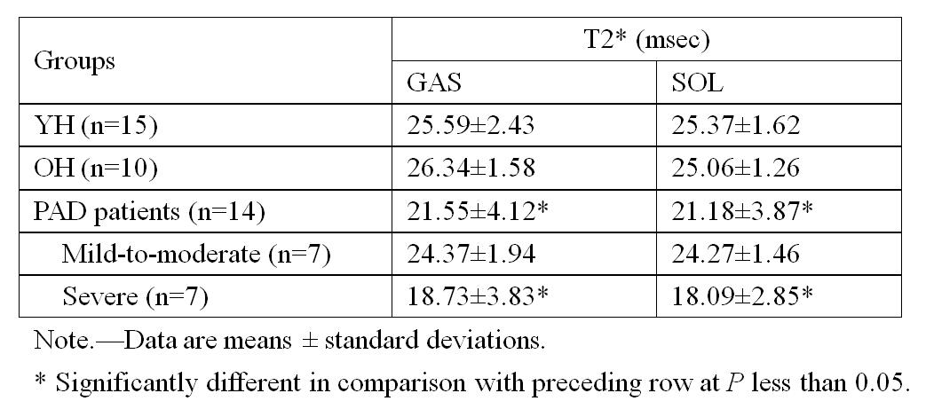
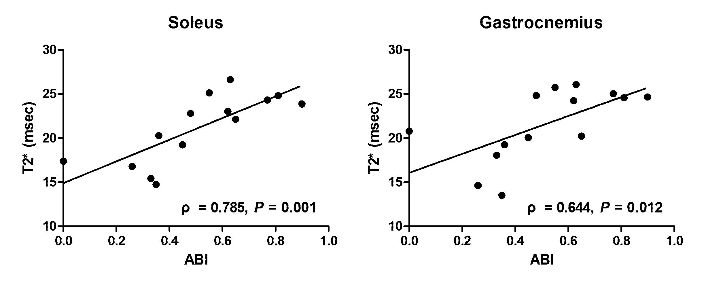
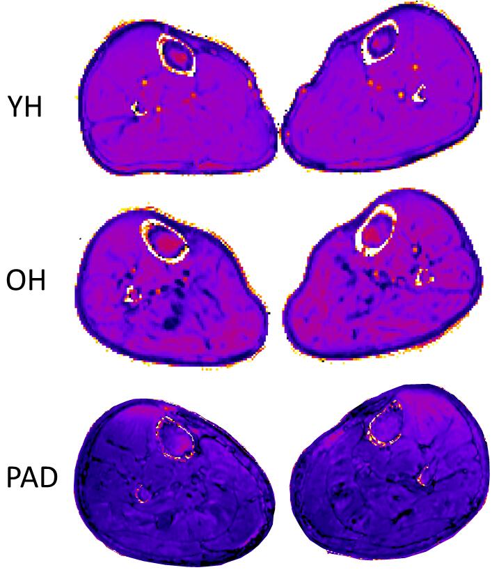
The local institutional review board approved the prospective study; written consent was obtained from all subjects. Fifteen healthy young subjects (YH), 14 patients with PAD, and 5 age-matched older healthy subjects (OH) were included. Healthy adults were recruited on the basis of their ankle-brachial index (ABI) >0.9 and no clinical evidence of PAD. Patients with PAD were recruited from the department of vascular surgery with symptoms of intermittent claudication, rest pain or critical limb ischemia and an ABI of ≤0.90 (0.50<ABI≤ 0.90, mild-to-moderate, n=7; ABI≤ 0.50, severe, n=7). All the MRI measurements were carried out on a 3.0-T MRI unit (HDxt, GE Healthcare, Waukesha, WI) with an eight-channel cardiac coil. Before BOLD MRI, axial three-dimensional spoiled gradient recalled echo T1-wegithed images were acquired for use as anatomic landmarks. BOLD was performed by using a multi-echo gradient recalled echo sequence implementing the following parameters: repetition time msec/echo times msec 875/(2.5, 6.7, 10.9, 15.1, 19.3, 23.5, 27.7, 31.9, 36.1, 40.3, 44.5, 48.7, 52.9, 57.1, 61.3, and 65.5); matrix, 256×128; FOV, 32×32 mm2; slice thickness/gap, 5/0 mm; number of slices, 12. BOLD data were post-processed on a pixel-by-pixel basis on a workstation (Advantage Workstation 4.5; GE Medical Systems) to obtain corresponding parameter maps of T2* relaxation time. Regions of interest (ROIs) were drawn manually in the gastrocnemius (GAS) and soleus (SOL) muscles on anatomical T1-weighted images and copied to corresponding BOLD maps. Mann-Whitney U test was performed to studied the T2* difference between each pair of groups (YH vs OH, OH vs PAD, and mild-to-moderate vs severe PAD). Correlation between T2* and ABI in PAD patients was assessed by using the Spearman rank correlation test. P-values <0.05 were considered as statistically significant.Results
T2* was found to be independent of age factor, with no significant difference observed between YH and OH in GAS and SOL (P = 0.567 and 0.531, respectively). T2* showed significant lower values in PAD patients compared to age-match OH, with P-values of 0.001 and 0.007 for GAS and SOL, respectively. In patient group, significant decreased T2* values were also demonstrated in severe PAD patients compared with mild-to-moderate PAD patients in GAS and SOL (P = 0.011 and 0.001, respectively). Spearman rank correlation analysis showed that, in PAD patients, T2* was significantly correlated with ABI in GAS (ρ = 0.644; P = 0.012) and SOL (ρ = 0.785; P = 0.001).Discussion and Conclusion
In our study, there were no significant differences between YH and OH for resting-state T2*, indicating that aging does not seem to affect BOLD in healthy subjects at rest. This finding was in accordance with other observations.3 However, lower T2* values were related to the presence of PAD and more disease severity stratified by ABI. With increasing ABI, the function of blood oxygenation in the capillary bed was declined, leading to a shortened T2* value. In a previous study, BOLD-derived metrics under the ischemia–reperfusion paradigm was also found to be correlated with ABI, suggestive of disease severity–dependent impairment of vascular response in PAD.4 Our results suggest that even at rest, vascular function at tissue level could be indicative of disease progression. In conclusion, our results showed that resting-state BOLD could be used as a useful technique to diagnose and assess the severity of PAD.Acknowledgements
This study was supported by the Shanghai Pujiang Program (grant 15PJ1405200) and National Natural Science Foundation of China (grants 81271638, 81501458 and 81571630).References
1. Jacobi B, Bongartz G, Partovi S, et al. Skeletal muscle BOLD MRI: from underlying physiological concepts to its usefulness in clinical conditions. J Magn Reson Imaging. 2012; 35:1253-1265.
2. Zuo C S, Sung Y H, Simonson D C, et al. Reduced T2* values in soleus muscle of patients with type 2 diabetes mellitus. PloS one, 2012; 7(11): e49337.
3. Kos S, Klarhöfer M, Aschwanden M, et al. Simultaneous dynamic blood oxygen level-dependent magnetic resonance imaging of foot and calf muscles: aging effects at ischemia and postocclusive hyperemia in healthy volunteers. Invest Radiol, 2009; 44(11): 741-747.
4. Englund EK, Langham MC, Ratcliffe SJ, et al. Multiparametric Assessment of Vascular Function in Peripheral Artery Disease Dynamic Measurement of Skeletal Muscle Perfusion, Blood-Oxygen-Level Dependent Signal, and Venous Oxygen Saturation. Circ Cardiovasc Imaging, 2015; 8:e002673.
Figures