5050
T2 mapping pseudo-color pictures and FS-FSE- PDWI in grading diagnosis of patellar cartilage damage:a retrospective study compared with arthroscopy1The first affiliated hospital of Dalian medical university, Dalian, China
Synopsis
Our institution use arthroscopy as the gold standard, apply T2mapping and FS-FSE- PDWI for the evaluation of patellar cartilage damage, to investigate value of T2 mapping pseudo-color pictures in assessment of patellar cartilage injury grading. This group of 45 cases of patellar cartilage damage, the correlations of T2mapping pseudo color grading,FS-FSE- PDWI grading and arthroscopic grading were analyzed, found the correlation between T2mapping pseudo color and arthroscopy precedes that between FS-FSE- PDWI and arthroscopy. Therefore, we believe that the T2mapping pseudo color can be used to evaluate the patellar cartilage damage, and better than FS-FSE- PDWI.
Purpose
T2 mapping MR as a new cartilage imaging technology, was previously mostly used for quantitative assessment of early cartilage damage(1). T2 value for quantitative assessment of different degrees of cartilage damage is still controversial. FS-FSE- PDWI has been proved to have high sensitivity, specificity and accuracy for cartilage lesions. Our institution use arthroscopy as the gold standard, apply T2mapping and FS-FSE- PDWI for the evaluation of patellar cartilage damage, to investigate value of T2 mapping pseudo-color pictures in assessment of patellar cartilage injury grading.
Materials and Methods
62 patients who underwent knee MR examination and arthroscopic surgery were collected, including 32 males and 30 females, aged 30-51 years, mean 40.7 years. GE Company Signa3.0T MR was used. The scan sequences include: FSE-T1WI, FSE-T2WI, FS-FSE-PDWI and T2mapping. T2mapping images were sent to the GE-ADW 4.3 workstation to generate T2 mapping pseudo-color pictures of patellar cartilage. ROIs were drawn consistent with arthroscopy. All MR images were reviewed by two radiologists for identify the ICRS grading of each patient. Arthroscopy surgeries were performed by two experienced specialists, using Outerbridge classification to classify cartilage damage degree. Spearman rank correlation test was used to evaluate correlation of T2 mapping pseudo-color pictures and FS-FSE-PDWI with arthroscopy. Rank sum test was used for comparing of cartilage damage grading in T2 mapping pseudo-color pictures and FS-FSE-PDWI.Results
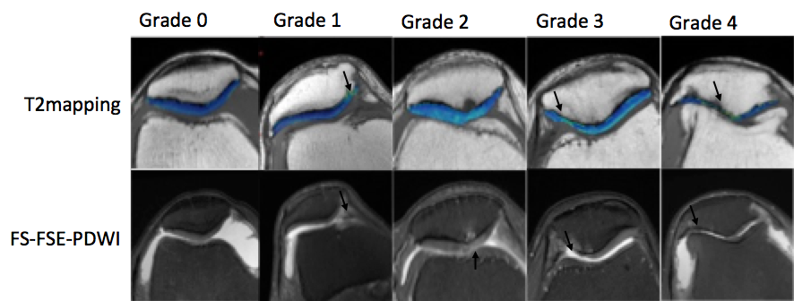
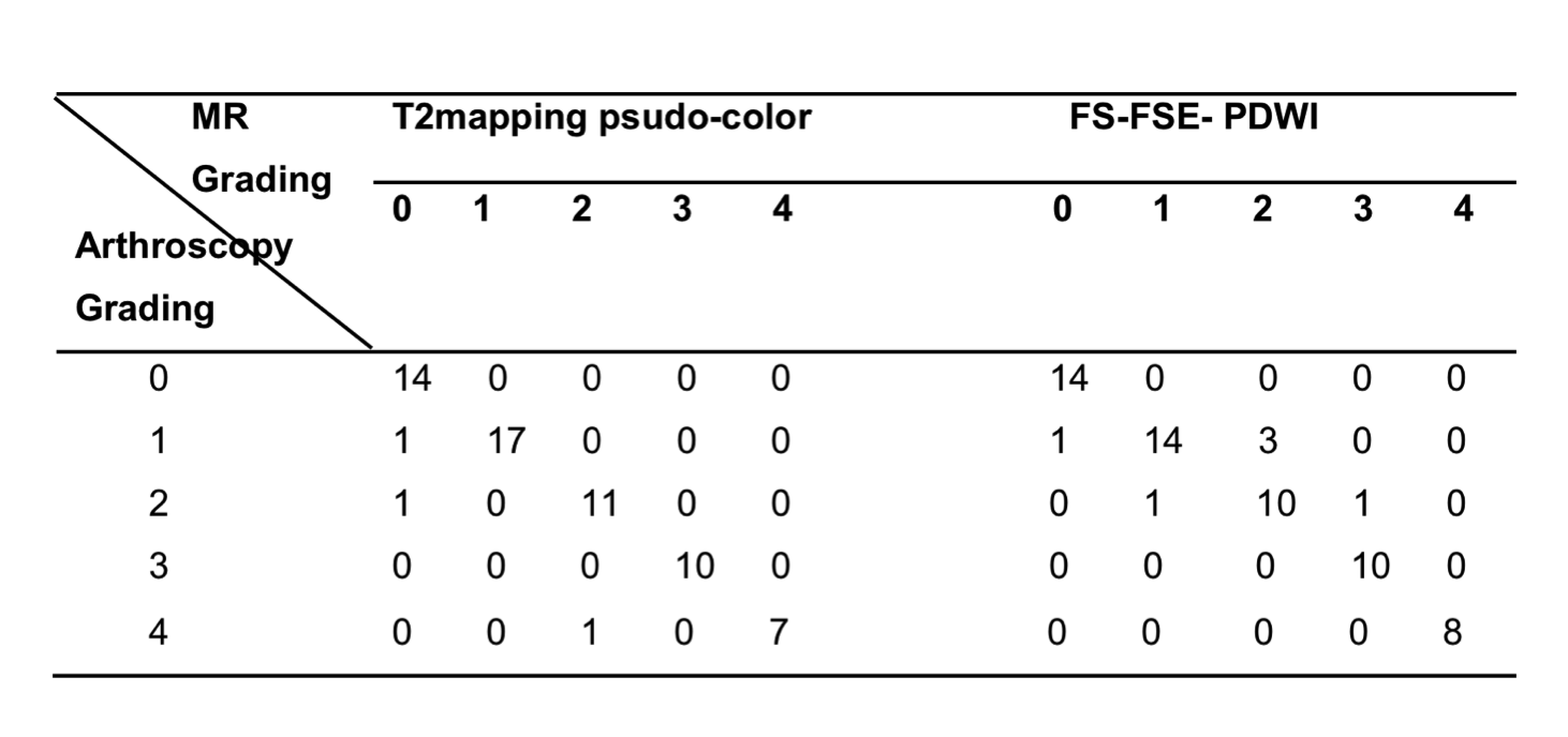
T2 mapping pseudo-color pictures showed a uniform blue color levels in normal patellar cartilage and blue levels of normal cartilage contrast with the gray levels joint effusion, which would be helpful to find cartilage defects. In 62 cases of patellar cartilage, 16 cases were diagnosed as normal patellar cartilage in T2 mapping pseudo-color pictures. I-IV grade injury were 17, 11, 10, 7 cases respectively. 14 cases were confirmed as normal patellar cartilage via arthroscopic surgery ,and I-IV grade injury were 18, 10, 10, 8 cases respectively. T2 mapping pseudo-color pictures and arthroscopy in the diagnosis of patellar cartilage damage of varying degrees of correlation coefficient rs = 0.956, P <0.05; the T2 mapping pseudo-color pictures and arthroscopic evaluation of cartilage damage difference test, p> 0.05, T2mapping pseudo-color pictures and arthroscopic damage to the evaluation of patellar cartilage grading was no significant difference.Discussion
In this study, we adjusted the threshold to make T2mapping pseudo color pictures showed a uniform blue color in normal patellar cartilage, gray levels near the joint effusion and green or yellow color in the lesions. In this condition the two layer of patellar cartilage depth resolution is poor, but good contrast between joint effusion and patellar cartilage can clearly show cartilage defect degree, so as to distinguish the degree of cartilage damage. This group of 45 cases of patellar cartilage damage, the correlations of T2mapping pseudo color grading,FS-FSE- PDWI grading and arthroscopic grading were analyzed, found the correlation between T2mapping pseudo color and arthroscopy precedes that between FS-FSE- PDWI and arthroscopy. Therefore, we believe that the T2mapping pseudo color can be used to evaluate the patellar cartilage damage, and better than FS-FSE- PDWI.Conclusion
T2 mapping pseudo-color pictures would be reliable in classification of patellar cartilage damage assessment.Acknowledgements
No acknowledgement found.References
1. S. Apprich, T.C. Mamischa TC, Welsch GH , et al. Quantitative T2 mapping of the patella at 3.0 T is sensitive to early cartilage degeneration, but also to loading of the knee. Eur J Radiol,2012,81(4):438-443Figures