5046
Comparison of DENSE-FISP and DENSE-FID for assessment of cartilage strain computation under compressive loading.1Human movement biomechanics research group, Department of kinesiology, KU Leuven, Leuven, Belgium, 2Biomedical MRI, KU Leuven, Leuven, Belgium, 3Department of Biomedical Engineering, Rensselaer Polytechnic Institute, Troy, NY, United States, 45. Department of Mechanical Engineering, University of Colorado Boulder, Boulder, CO, United States
Synopsis
While true-FISP provides increased signal to noise ratio, it is also prone to banding artefacts due to incoherent dephasing. We thus here compare true-FISP DENSE with FID-DENSE to estimate cartilage strain under compressive loading. As FID-DENSE will be artefact free however at the expense of a lower signal to noise ratio, we investigate if DENSE-FID or DENSE-FISP will provide a more robust computation of cartilage strains under compressive loading.
Purpose
Assessment of mechanical properties of healthy and diseased articular cartilage is of particular interest. We have recently shown that cartilage displacement and strain metrics were correlated to global molecular changes assessed through T1, T2 and T1rho (1). Therefore, non-invasive monitoring of cartilage deformation can potentially serve as a unique biomarker for tissue damage and disease progression. DENSE-FISP is quite often preferred for in-vivo assessment due to the improved signal to noise but is also prone to banding artefacts (2). The aim of this study was thus to investigate if either DENSE-FID or DENSE-FISP will provide the best option for the computation of cartilage strain under compressive loading.Methods
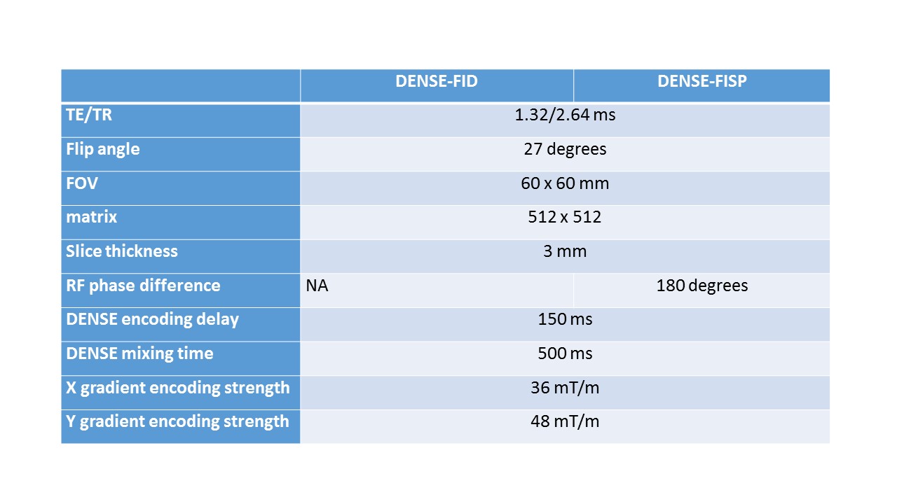
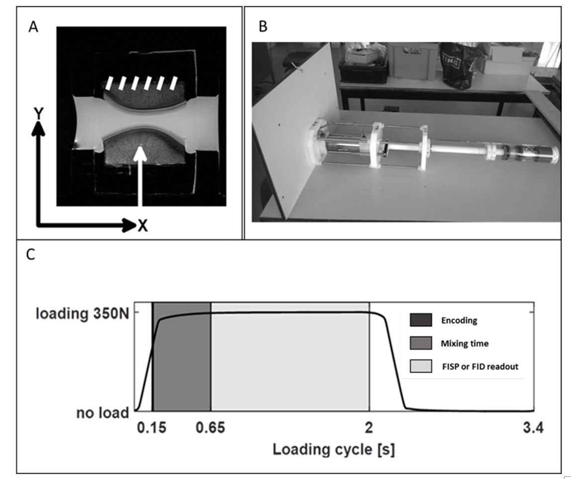
Eight fresh knee joints (9 month old bovine) were used. Osteochondral explants with 30mm diameter were harvested from load-bearing area of medial and lateral femoral condyles and mounted on a custom-built MRI compatible loading device. All MRI data were acquired using a 9.4T MRI system (Bruker Biospec 94/20USR) with a quadrature transmit/receive coil (72mm internal diameter). Before each scan session, a 2D T2 weighted RARE reference was acquired (TEeff/TR:16.9/5692 ms, RARE factor:4, 9 contiguous slices with 3 mm thickness). Before image acquisition during loading, the osteochondral plugs were cyclically loaded for ~500 cycles to reach quasi-steady state load-deformation behavior (cycle of 2.0 s constant loading with 350N and 1.4 s unloading). To image the displacements under compressive loading, DENSE sequences with both FISP and FID readouts were used (see Table 1 for sequence details). Three consecutives scans were acquired with no displacement encoding in any direction (reference scan), 36 mT/m gradient encoding in X-direction and 48 mT/m in Y (Fig.1). For each sample, signal to noise ratio (SNR) were determined. Phase derivative variance maps were computed to assess the quality of the phase image before unwrapping and efficiency of automatic phase unwrapping was assessed by computing the percentage of residual voxels eliciting variations higher than π to the surrounding voxels. Finally, compressive (Exx), tangential (Eyy) and shear (Exy) strains from DENSE-FISP and DENSE-FID were compared through Blant-Altman plots.Results
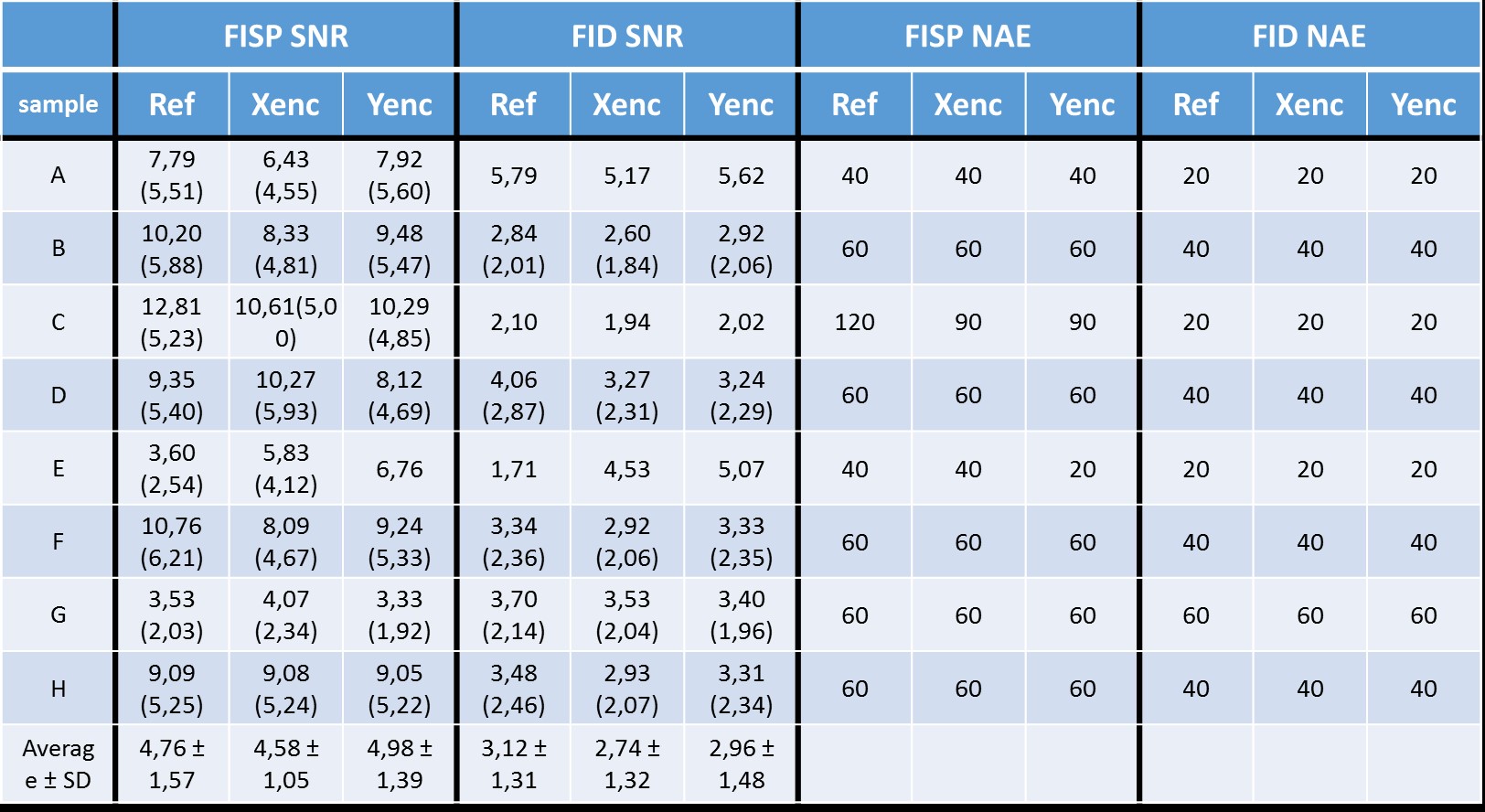
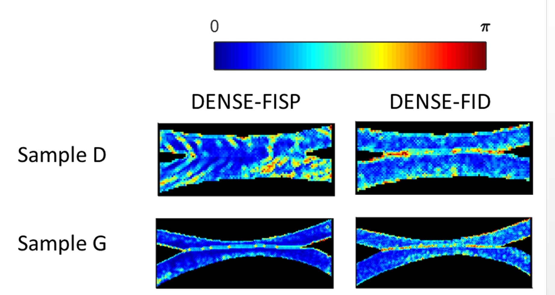
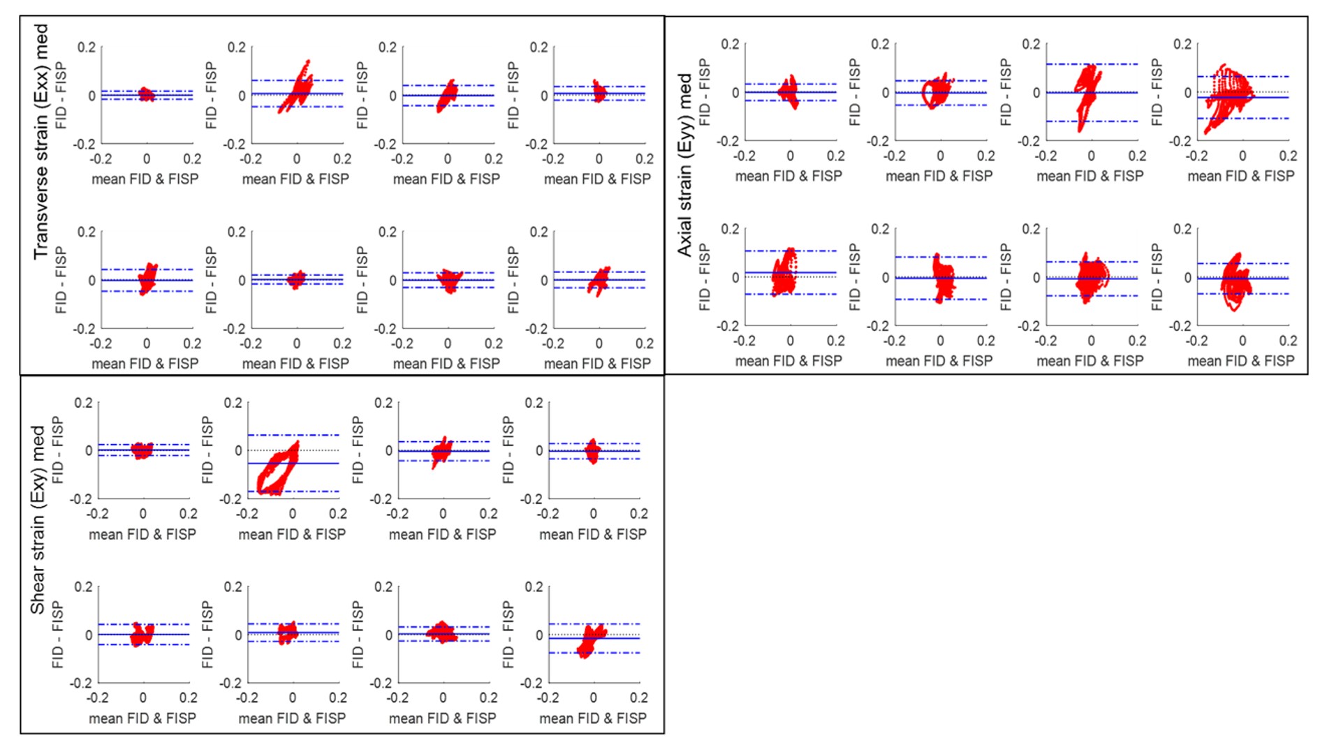
Signal to noise ratio for the different conditions are summarized in Table2. DENSE-FISP has an overall SNR of 4.77 ± 1.30 compared to 2.94 ± 1.45 for FID for 20 averages. The phase derivative variance maps before the unwrapping showed an overall good homogeneity for FISP. Nevertheless, in some samples banding artefacts are clearly visible (Fig.2). In contrast, phase information from FID was noisier but remained artefact free. The residue of pixels exceeding π to the neighbors’ voxels after automatic phase unwrapping was higher for FID data (4.68 ± 5.27% vs 14.47 ± 12.95% for FISP and FID respectively), indicative of efficient automatic unwrapping for the FISP data. Overall the Bland-Altman plots (fig. 3) demonstrated that both methods were in agreement with only few outliers for the computation of Exx, Eyy and Exy from the contact region.Discussion and conclusion
Overall FISP readout provides data of higher quality through improved SNR which is also reflected by a higher homogeneity of the phase information. However, in 10-20% of the samples, the banding artefacts due to off-resonance effect, may corrupt the data rendering them unusable. This highlight the importance of good shimming which may be more difficult at high field or in situations with lower field homogeneiity including implants and air pockets.Acknowledgements
This research was supported in part by a Ph.D. grant of the Agency for Innovation by Science and Technology (IWT) 131105, the KU Leuven research council OT13083 and IMIR PF10/0117, HERCULES AKUL/13/29 and NIH R01 AR063712 and R21 AR064178.References
1. Willy Gsell, Willy Zevenbergen, Tom Dresselaers, Deva Chan, Corey Neu, Uwe Himmelreich, and Ilse Jonkers. Biomechanical properties of bovine knee cartilage under compressive loading: A study at high field MRI (9.4T) using T1, T2 and T1rho relaxometry combined with DENSE-FID. Proceedings of the 25th Annual Meeting of ISMRM, Honolulu, USA. April 22-27 2017, abstract 1147.
2. Xiang QS, Hoff MN. Banding artifact removal for bSSFP imaging with an elliptical signal model. Magn Reson Med. 2014 Mar;71(3):927-33.
Figures