5043
3D Ultrashort Echo Time MRI for Evaluation of Cartilaginous Endplate of Lumbar Disc In Vivo: Feasibility and Correlation with Disc Degeneration in T2 weighted Spin Echo Sequence1Radiology, Inha University Hospital, Incheon, Republic of Korea, 2Soonchunhyang University Bucheon Hospital, Bucheon, Republic of Korea
Synopsis
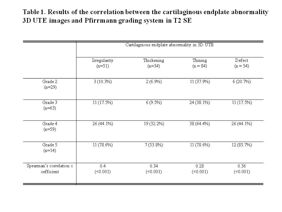
Total 165 discs of 33 patients were imaged with sagittal 3D UTE (TR = 16.1 ms, TE = 0.032 ms and 6.6 ms, echo-subtraction) with 3D cones trajectory technique and conventional sagittal T2 weighted (TR = 3232, TE = 91.4) SE in 3T MRI. Two musculoskeletal radiologists evaluated the CEP abnormalities such as irregularity, thickening, thining, and defects in the 3D UTE with consensus and correlated with the Pfirrmann grading system in the sagittal T2 SE. In result, all of the CEP abnormalities were positively correlated with the Pfirrmann grading system with statistical significance (p < 0.001).
Synopsis
Total 165 discs of 33 patients were imaged with sagittal 3D UTE (TR = 16.1 ms, TE = 0.032 ms and 6.6 ms, echo-subtraction) with 3D cones trajectory technique and conventional sagittal T2 weighted (TR = 3232, TE = 91.4) SE in 3T MRI. Two musculoskeletal radiologists evaluated the CEP abnormalities such as irregularity, thickening, thining, and defects in the 3D UTE with consensus and correlated with the Pfirrmann grading system in the sagittal T2 SE. In result, all of the CEP abnormalities were positively correlated with the Pfirrmann grading system with statistical significance (p < 0.001).Background
Identifying changes in cartilaginous endplate (CEP) morphology may be important for the diagnosis and prognosis of degenerative disc disease (1) However, little is known about the MRI characteristics of changes in CEP morphology, as the cartilage in the CEP has short T2 relaxation time values, wherein its signal is not captured in conventional MRI sequences using long echo times (2). Recently, Bae et al. (3) reported that MRI sequences with very short echo times, referred to as ultrashort echo time (UTE) sequences, enabled direct visualization of uncalcified and calcified CEPs. However, the clinical utility of UTE for degenerative disc diseases in vivo is still unknown. The purpose of our study was to investigate the association between CEP abnormalities and disc degeneration in the T2-weighted spin-echo sequence (T2 SE) in lumbar discs in vivo.Methods
Total 165 discs of 33 patients ( 16 men, 17 women, mean age: 58.9 year old, age range: 21- 81 year old) were imaged with sagittal 3D UTE (TR = 16.1 ms, TE = 0.032 ms and 6.6 ms, echo-subtraction) with 3D cones trajectory technique and conventional sagittal T2 weighted (TR = 3232, TE = 91.4) SE in 3T MRI. Two musculoskeletal radiologists evaluated the CEP abnormalities such as irregularity, thickening, thining, and defects in the 3D UTE with consensus and correlated with the Pfirrmann grading system (4) in the sagittal T2 SE using Pearson’s chi-square test and Spearman’s correlation analysis.Results
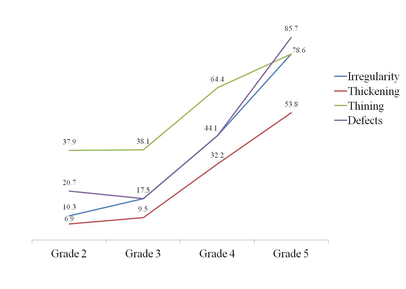
The frequency of CEP abnormality and grades of disc degenerations were in table 1. In 3D UTE, most common CEP abnormality was thining. In conventional T2 SE, grade 3 disc degeneration was most common. None of the disc showed grade 1. All of the CEP abnormalities were positively correlated with the Pfirrmann grading system with statistical significance (p < 0.001 for both Pearson’s chi-square and Spearman’s correlation analysis, table 1, figure 1). However, the strength of correlations was weak except the irregularity (table 1).Discussion
The structural and micromechnical change of CEP reduces the diffusion of nutrients to the cells of the NP, and leads to decreased proteoglycan content within the disc, disc calcification, fibrosis and rupture. Therefore, identifying changes in CEP morphology may be important for the diagnosis and prognosis of degenerative disc disease. In our study, the CEP abnormalities in 3D UTE positively correlated with the severity of disc degeneration in conventional SE. This may reflect the association between CEP abnormality and disc degeneration corresponding with prior studies (5, 6). However, the weak correlation between the CEP abnormalities and the Pfirrmann grading system was somewhat disappointing. The signal intensity and disc space in T2 SE images depend on the proteoglycan levels, water content, and hydrostatic pressure of the disc [7, 8]. Changes in the proteoglycan and hydration levels, known as biochemical aging, begin at birth, mainly in the NP (9). Structural changes in CEPs tend to appear later, usually from 20 years old (10). The results may indicate that biochemical aging occurs to some extent before any structural CEP changes. In addition, the CEP had only a submillimeter structure, which requires high image quality for accurate visualization (11). Due to the low image quality of the 3D UTE, the subtle CEP abnormalities might not have been detected and vice versa and normal CEPs might have been misinterpreted as abnormal. Despite these limitations, we cautiously expect that the detection of early CEP abnormality afforded by 3D UTE might provide a non-invasive comprehensive evaluation of early disc degeneration and its pathophysiology in the future.Conclusions
The CEP abnormality in 3D UTE MRI may be associated with the severity of T2 SE disc degeneration.Acknowledgements
No acknowledgement found.References
1. Bogduk N, Endres SM. Clinical anatomy of the lumbar spine and sacrum. New York: Elsevier/Churchill Livingstone, 2005
2. Lotz JC, Fields AJ, Liebenberg EC. The role of the vertebral endplate in low back pain. Global Spine J 2013; 3:153-164
3. Bae WC, Statum S, Zhang Z et al. Morphology of the cartilaginous endplates in human intervertebral disks with ultrashort echo time MR imaging. Radiology 2013; 266:564-574
4. Pfirrmann CW, Metzdorf A, Zanetti M, Hodler J, Boos N. Magnetic resonance classification of lumbar intervertebral disc degeneration. Spine (Phila Pa 1976). 2001;26(17):1873-8.
5. Bian Q, Liang QQ, Wan C et al. Prolonged upright posture induces calcified hypertrophy in the cartilage end plate in rat lumbar spine. Spine 2011; 36:2011-2020
6. Ariga K, Miyamoto S, Nakase T et al. The relationship between apoptosis of endplate chondrocytes and aging and degeneration of the intervertebral disc. Spine (Phila Pa 1976) 2001; 26:2414-2420
7. Modic MT, Pavlicek W, Weinstein MA et al. Magnetic resonance imaging of intervertebral disk disease. Clinical and pulse sequence considerations. Radiology 1984; 152:103-111
8. Modic MT, Ross JS. Lumbar degenerative disk disease. Radiology 2007; 245:43-61
9. Roberts S, Evans H, Trivedi J, Menage J. Histology and pathology of the human intervertebral disc. J Bone Joint Surg Am 2006; 88 Suppl 2:10-14
10. Adams MA, Freeman BJ, Morrison HP, Nelson IW, Dolan P. Mechanical initiation of intervertebral disc degeneration. Spine (Phila Pa 1976) 2000; 25:1625-1636
11. Nerlich AG, Schleicher ED, Boos N. 1997 Volvo Award winner in basic science studies. Immunohistologic markers for age-related changes of human lumbar intervertebral discs. Spine (Phila Pa 1976) 1997; 22:2781-2795
Figures