5038
Compositional assessment of low-grade cartilage lesions using T2 mapping at 3 and 7 Tesla MRI: a one year follow-up study1Department of Biomedical Imaging and Image-Guided Therapy, Medical University of Vienna, Vienna, Austria, 2Department of Imaging Methods, Institute of Measurement Science, Bratislava, Slovakia, 3Department of Orthopedics, Medical University of Vienna, Vienna, Austria, 4Novartis Institutes for Biomedical Research, Basel, Switzerland, 5Research Unit of Medical Imaging, Physics and Technology, University of Oulu, Oulu, Finland, 6Department of Traumatology, Medical University of Vienna, Vienna, Austria, 7Division of Radiological Physics, Department of Radiology, University of Basel Hospital, Basel, Switzerland, 8Slovak Academy of Sciences, Institute of Measurement Science, Bratislava 4, Slovakia, 9Christian Doppler Laboratory for Clinical Molecular MR Imaging, Vienna, Austria
Synopsis
T2 maps were assessed as a potential marker for the long-term follow-up of the patients with cartilage lesions ICRS Grade I-II in five time points (baseline, 8 days, 3, 6 and 12 months). For the T2 mapping, a 3D triple echo steady state sequence which is capable of delivering high quality high-resolved T2 maps at ultra-high field MRI was used. We observed a significant decrease in T2 values at 3T over time in superficial zone of the cartilage defect. There was no statistically significant change at 7T. T2 mapping could be used in the future as a good alternative to cartilage biopsies in clinical trials on new therapies aimed at cartilage regeneration.
Background & Purpose
T2 mapping has been previously demonstrated as a reliable quantitative method for assessing the collagen network damage and water content in human articular cartilage 1,2. The sensitivity and reliability of T2 mapping for subtle cartilage lesions at different fields have not yet been sufficiently investigated. The purpose of this study was to investigate MRI T2 mapping as a possible marker to follow-up of the patients up to one year after cartilage injury and to compare the reliability at high field (3T) and ultra-high field (7T).Methods and Materials:
Twenty-one patients with femoral cartilage defect(s) in the knee joint after traumatic injury with ICRS Grade I and II (mean age, 46.3/11.1 years; 12 males/9 females) were examined. T2 maps were acquired for each of these patients on two MR scanners: 3T Prisma Fit (Siemens, Erlangen, Germany) and 7T Magnetom scanner (Siemens Healthcare, Germany). T2 relaxation times were calculated using a 3D-Triple Echo Steady State sequence (3D-TESS)3. Regions of interest (ROIs) were defined on morphological images in the region suspicious for a cartilage defect and in a region of normal-appearing cartilage (weight- and non-weight bearing region) in both deep and superficial cartilage layers and transferred onto T2 maps. The measurements were repeated at five time-points (baseline, 8 days, 3 months, 6 months and 12 month after baseline). Student’s t-test was used to determine the potential differences in T2 over time at different field strengths.Results
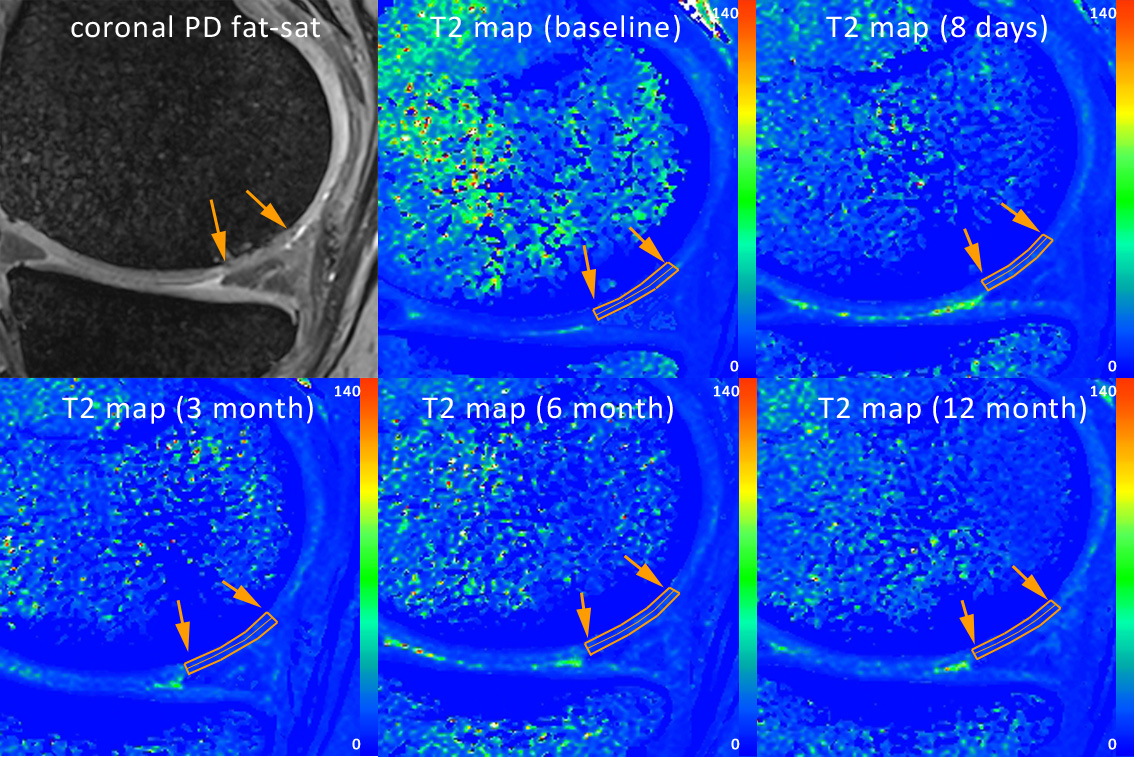
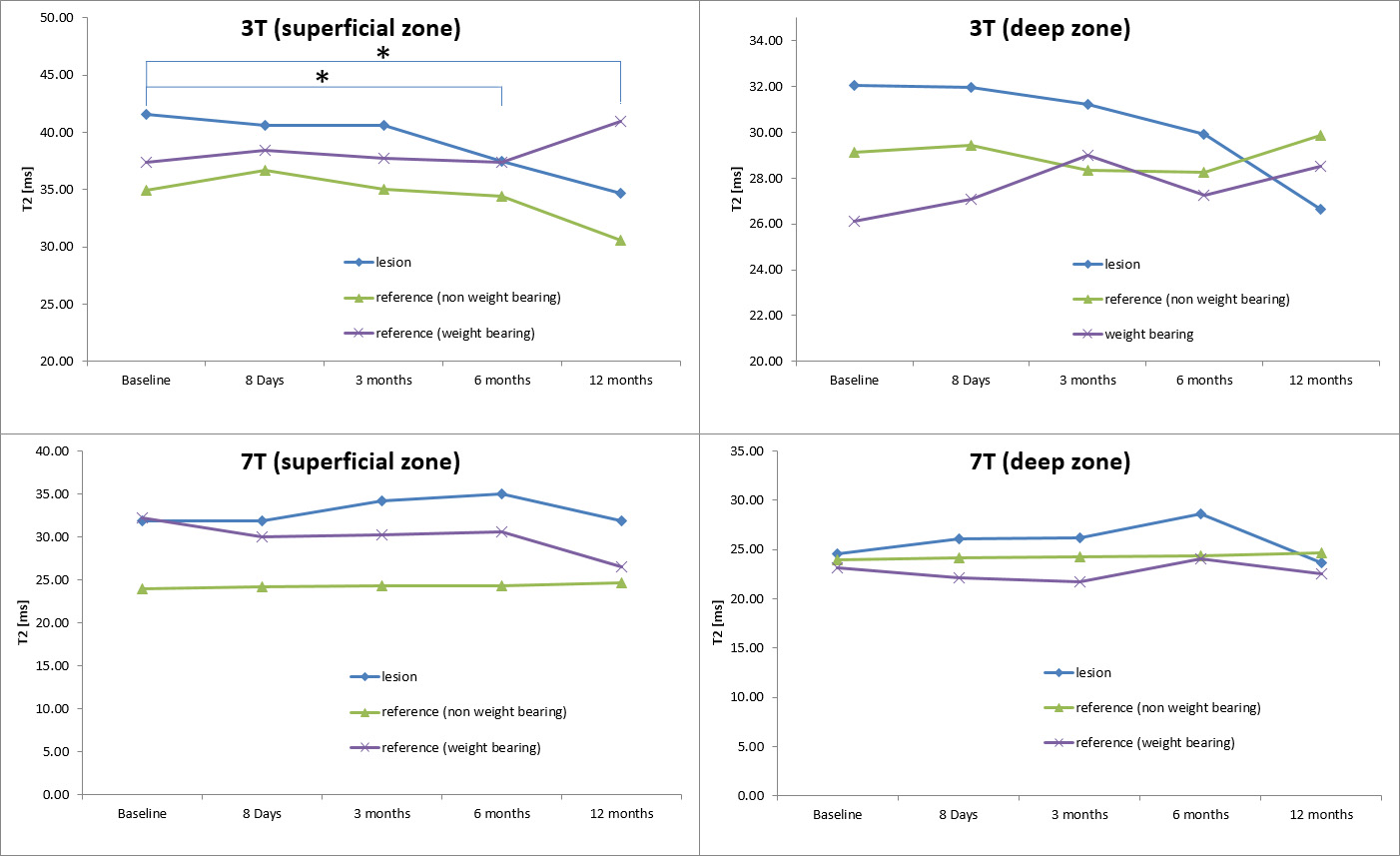
Illustrative T2 maps with the ROI position are shown on Fig. 1. We observed a decrease of T2 values at 3T, whilst the T2 values at 7T did not change. At 3T we found a statistical significant drop of T2 in the superficial layer of the defect between baseline and 6 month follow-up: 41.85±8.57ms to 36.21±7.15ms (change of 5.64±1.41ms, p = 0.0321) and again in the superficial layer between baseline and 12 month follow-up: 41.85±8.57ms to 35.28±4.93ms (change of 6.57±3.63ms, p = 0.0381). No statistical significance was found in 7T T2 values. The T2 values progress is visualized in Fig. 2.Conclusion
T2 mapping with 3D-TESS enables monitoring of biochemical changes in low-grade cartilage lesions over time. It seems that T2 values acquired at 3T are more sensitive to collagen network because of an echo time of ~4ms and the resulting contribution of short T2 component to the signal which is already decayed at 7T. Hence, T2 may be considered as a valuable marker for monitoring cartilage development after traumatic injury causing alterations to the collagen matrix or/and regenerative therapy.Acknowledgements
This work was supported by the Slovak Research and Development Agency: APVV-15-0029, Grant Agency of the Slovak Academy of Sciences (VEGA 2/0001/17) and Austrian Science Fund (FWF) KLI541-B30.References
[1] Mosher TJ at al. Cartilage MRI T2 relaxation time mapping: overview and applications, Semin Musculoskelet Radiol 2004; 08(4): 355-368
[2] Liess, C at al. Detection of changes in cartilage water content using MRI T2-mapping in vivo, Osteoarthritis and Cartilage 2002, 10(12): 907-913
[3] Heule R et al. Triple echo steady-state (TESS) relaxometry, Magnetic Resonance in Medicine 2004; 71(1) 230–237
Figures