5034
Depth dependence of diffusion in articular cartilage of the knee visualized at 7T1Radiology, University Medical Center Utrecht, Utrecht, Netherlands, 2Radiology, Academic Medical Center, Amsterdam, Netherlands, 3Biomedical Engineering and Physics, Academic Medical Center, Amsterdam, Netherlands, 4Biomedical Engineering, Eindhoven University of Technology, Eindhoven, Netherlands, 5Orthopaedics, University Medical Center Utrecht, Utrecht, Netherlands
Synopsis
The goal of this study was to apply high-resolution diffusion weighted imaging (DWI) at 7T to assess depth dependence of diffusion in the articular cartilage in the knee. Four healthy volunteers were scanned with a diffusion prepared TSE sequence. ADC maps were consequently used to assess the depth dependence of ADC throughout the articular cartilage. The ADC value was shown to significantly increase from the bone-cartilage interface to the superficial zone. This work shows that the significant depth dependence of the ADC in cartilage layers can be observed in vivo on 7T with diffusion weighed imaging.
Introduction
Early cartilage damage results in changes to the orientation of collagen and the proteoglycan content, mostly in the superficial zone. Thus, identifying zonal changes would be important for the assessment of cartilage damage [1, 2]. In a previous 3T study of cartilage repair patients, zonal differences in cartilage were observed, but were not shown to be significant [3]. To better measure such differences in cartilage zones, a high resolution imaging protocol is needed because of the limited thickness of cartilage, which is about 2 to 4 mm depending on the location in the knee. Therefore, the purpose of this study was to apply high-resolution diffusion weighted imaging (DWI) at 7T to assess depth dependence of diffusion in the articular cartilage in the intact knee.Methods
Four subjects, without known pathology, were scanned on a 7.0T whole body scanner (Achieva; Philips Healthcare, Best, Netherlands) using a birdcage volume transmit coil with a dedicated receive array. A diffusion-prepared stimulated-echo turbo spin echo (DPsti-TSE) sequence [4] was implemented with the following imaging parameters: sagittal field of view (FOV) = 160*160*48 mm3, voxel size = 0.7 × 0.7 × 3.0 mm3, shot interval = 2000 ms, echo train length = 20, echo spacing = 38 ms, number of signal averages (NSA) = 2, SENSE = 3(AP) and 2(RL), b = 0, 400, total acquisition time = 5 min and 2 seconds. Post-processing was carried out by registering b400 images to b0 images using an affine method and fitting the data to the model
S(b) = S0 ∙ exp-b∙ ADC
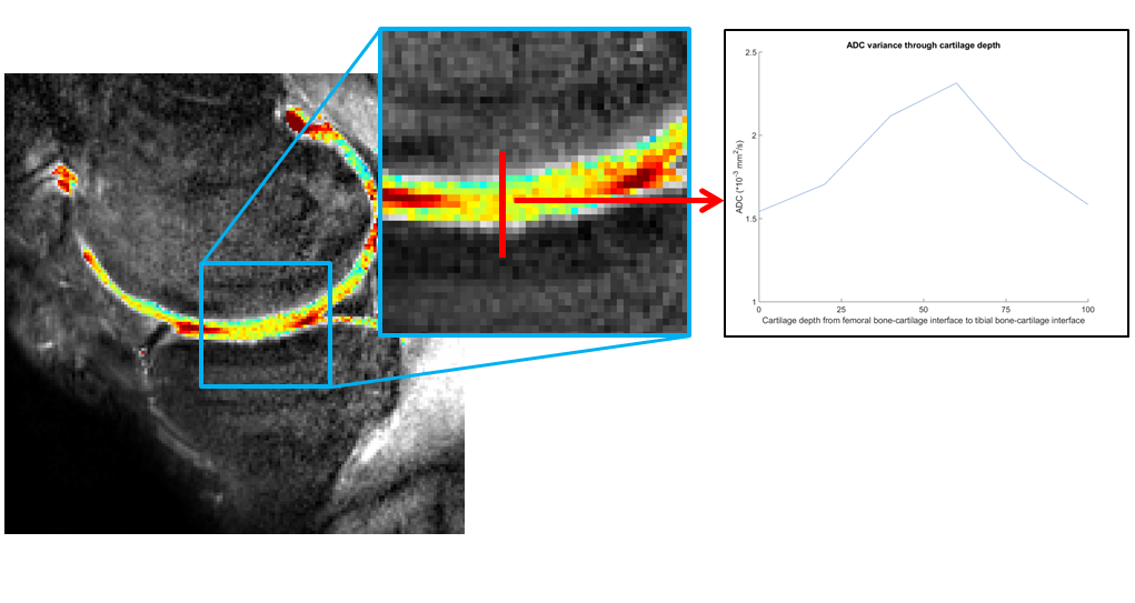
where S(b) is the diffusion weighted images, S0 is the non-diffusion weighted images, ADC is the desired apparent diffusion coefficient in mm2/s and b is the diffusion weighting strength in s/mm2. Depth dependence was assessed in the articular cartilage of the femur vs. that in the tibia. The ADC was measured in a large (between 15 and 20) number of profiles through the articular cartilage, perpendicular to the articular surface. One profile consists of a line of voxels from the bone-cartilage interface of the femoral cartilage, to the superficial zone of the femoral cartilage and the superficial zone of the tibial cartilage and ending with bone-cartilage interface of the tibia. An example of one profile and the way its retrieved is shown in figure 1. The mean profiles were calculated per volunteer to assess the depth dependence of ADC values throughout the cartilage thickness. Statistical significance of this depth dependence of ADC values throughout the cartilage was assessed with a repeated measures ANOVA.
Results
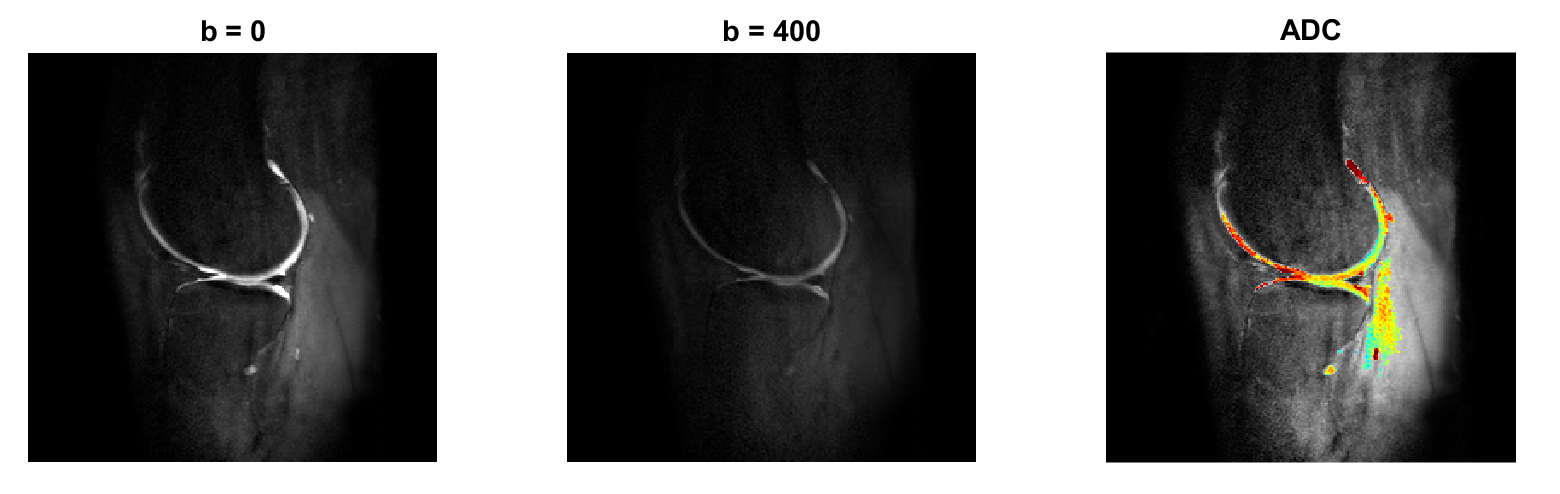
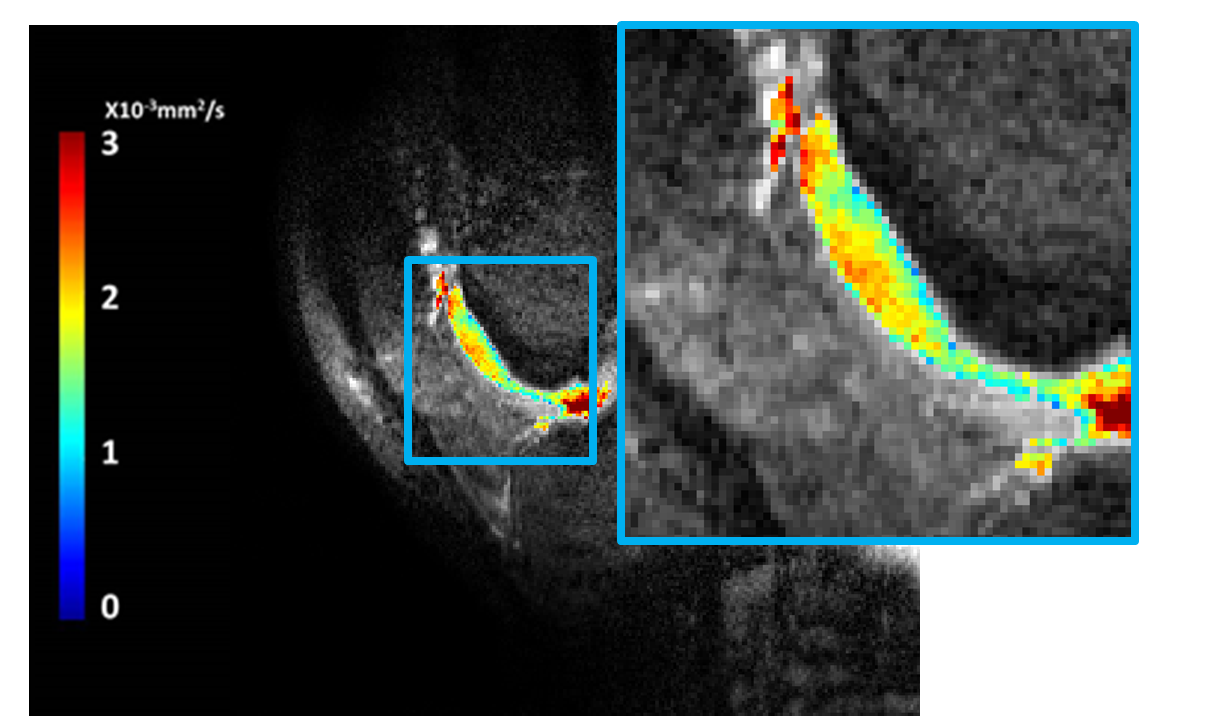
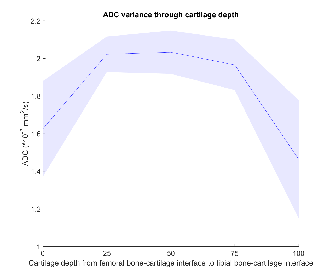
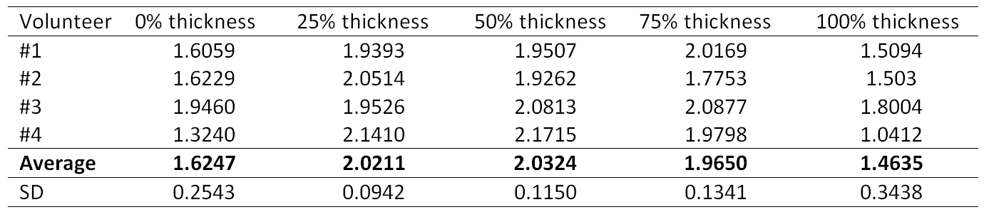
MRI acquisition was successful in all participants. Example images of the b=0 and b=400 images are shown in figure 2 (left and middle) for femoral and tibial cartilage. Figure 2 (right) shows the overlay of the calculated ADC values. One typical example of depth dependence can be seen on figure 3, which shows the ADC gradient in the trochlear groove. The depth dependence of ADC values over the profiles is shown in figure 4. It can be observed that the ADC value is lower on the bone-cartilage interfaces (0 and 100% in this graph) and higher on the superficial zones of the cartilage (50% in this graph). The difference in ADC values throughout the depth of the cartilage was shown to be significant (p = 0.003). The ADC values of the mean profile of each volunteer is given in table 1 with corresponding standard deviation.Discussion
Within this work we were able to show a significant depth dependence of ADC throughout the thickness of cartilage in vivo. These differences match with previous in vitro work, which also showed an increase of ADC from the bone-cartilage interface to the superficial zone [5–7]. Using this diffusion prepared stimulated echo approach in combination with higher SENSE acceleration factors, we were able to decrease the acquisition time from 14 minutes to 5 minutes as compared with the similar high resolution shown previously on 7T by Raya and colleagues [8]. However, with the limited subjects included, we could not yet observe a significant ADC difference per layer, which would further stimulate the design of even higher spatial resolutions.Conclusion
This work shows that a significant depth dependence of the ADC in cartilage layers can be observed in vivo on 7T with diffusion weighed imaging.Acknowledgements
No acknowledgement found.References
1. Saarakkala S, Julkunen P, Kiviranta P, et al (2010) Depth-wise progression of osteoarthritis in human articular cartilage: investigation of composition, structure and biomechanics. Osteoarthritis Cartilage 18:73–81. doi: 10.1016/j.joca.2009.08.003
2. Deng X, Farley M, Nieminen MT, et al (2007) Diffusion tensor imaging of native and degenerated human articular cartilage. Magn Reson Imaging 25:168–171. doi: 10.1016/j.mri.2006.10.015
3. Friedrich KM, Mamisch TC, Plank C, et al (2010) Diffusion-weighted imaging for the follow-up of patients after matrix-associated autologous chondrocyte transplantation. Eur J Radiol 73:622–628. doi: 10.1016/j.ejrad.2008.12.017
4. Zhang Q, Coolen BF, Versluis MJ, et al (2017) Diffusion-prepared stimulated-echo turbo spin echo (DPsti-TSE): An eddy current-insensitive sequence for three-dimensional high-resolution and undistorted diffusion-weighted imaging. NMR Biomed 30:1–12. doi: 10.1002/nbm.3719
5. Filidoro L, Dietrich O, Weber J, et al (2005) High-resolution diffusion tensor imaging of human patellar cartilage: Feasibility and preliminary findings. Magn Reson Med 53:993–998. doi: 10.1002/mrm.20469
6. Meder R, de Visser SK, Bowden JC, et al (2006) Diffusion tensor imaging of articular cartilage as a measure of tissue microstructure. Osteoarthr Cartil 14:875–881. doi: 10.1016/j.joca.2006.03.002
7. Mlynárik V, Sulzbacher I, Bittšanský M, et al (2003) Investigation of apparent diffusion constant as an indicator of early degenerative disease in articular cartilage. J Magn Reson Imaging 17:440–444. doi: 10.1002/jmri.10276
8. Raya JG, Horng A, Dietrich O, et al (2012) Articular cartilage: in vivo diffusion-tensor imaging. Radiology 262:550–559. doi: 10.1148/radiol.11110821
Figures