4991
High resolution frequency difference mapping using ASPIRE phase combination1High Field Magnetic Resonance Centre, Department of Biomedical Imaging and Image-guided Therapy, Medical University of Vienna, Vienna, Austria
Synopsis
Frequency difference mapping (FDM) is a promising new method for investigating tissue microstructure. To date, images have been of quite low resolution and noise because of the need to use monopolar readout and two difference operations. We propose an improved approach – ASPIRE-FDM – which is based on a recently developed coil combination method which removes both phase offsets and phase gradients in readout direction in more efficient bipolar acquisitions. The resulting maps have drastically reduced noise and make feasible a four-fold reduction in voxel volume compared to prior work.
Purpose
To improve the resolution and contrast of Frequency Difference Mapping (FDM) [1,2] by using time-efficient bipolar GRE acquisitions and optimized phase combination and processing.Theory
The non-linear phase evolution that is analyzed in FDM generates contrast related to the tissue microstructure and orientation to B0 [1]. The FDM calculation requires two phase subtraction steps: the first removes time-independent phase offsets and the second removes linear and non-local phase evolution [2]. Noise in the resulting FDM has been compensated for by using averaging (e.g. 10 repetitions in [2]).
The first phase subtraction step can be omitted if the ASPIRE [3] method of combining coil signals is used, as this eliminates phase offsets. The remaining phase subtraction step in the improved version, ASPIRE-FDM, is formulated in complex as
$$FDM(TE_n)=S(TE_n)\cdot{conj}(S(TE_1)^n)$$
ASPIRE also allows bipolar (rather than, to date, monopolar) data acquisition, as linear readout-gradient-related phase is removed. In a monopolar acquisition, strong “flyback” gradients are required between echoes, which are all acquired with the same readout gradient polarity. Bipolar acquisitions acquire echoes with both gradient polarities, avoiding the need for flyback gradients, making them more efficient and allowing increased resolution.
Methods
Data was acquired from a healthy volunteer with a 7T MR whole body Siemens MAGNETOM scanner with a 32-channel Nova Medical head coil.
Scan 1 was acquired according to Ref. [1] to compare the original FDM method with ASPIRE-FDM and Scan 2 with the higher resolution possible in a bipolar acquisition. Both were 3D multi-echo GRE scans with a field of view of 224 mm x 224 mm x 52 mm, GRAPPA 2 and slice and phase partial Fourier factors of 6/8. No averages were used, in contrast to Ref. [2] (where the number of averages was 10), with a view to approaching a clinically feasible acquisition time.
Scan 1: - a bipolar acquisition with 16 echoes spaced by 2 ms and starting at 2 ms. For the original FDM calculation it is effectively used as a monopolar acquisition [1], as only the 8 even echoes spaced 4 ms were used to avoid effects from different readout polarity. The same approach is taken here for the original FDMs. The receiver bandwidth was 698 Hertz/pixel, the voxel size was isotropic 1 mm and the acquisition time 4 m 9 s.
Scan 2: - a bipolar acquisition with the same 8 echo times as the even echoes in Scan 1. The resolution was doubled in readout and phase encode direction to yield a voxel size of 0.5 mm x 0.5 mm x 1 mm and the receiver bandwidth lowered to 272 Hertz/pixel with a slightly longer acquisition time of 4 m 25 s.
Results
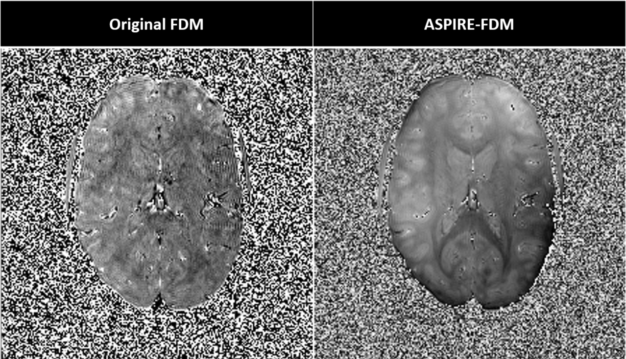
In Fig. 1, the original FDM image has high noise with hardly visible structures. The images calculated with ASPIRE-FDM have lower noise and high contrast between white and grey matter.
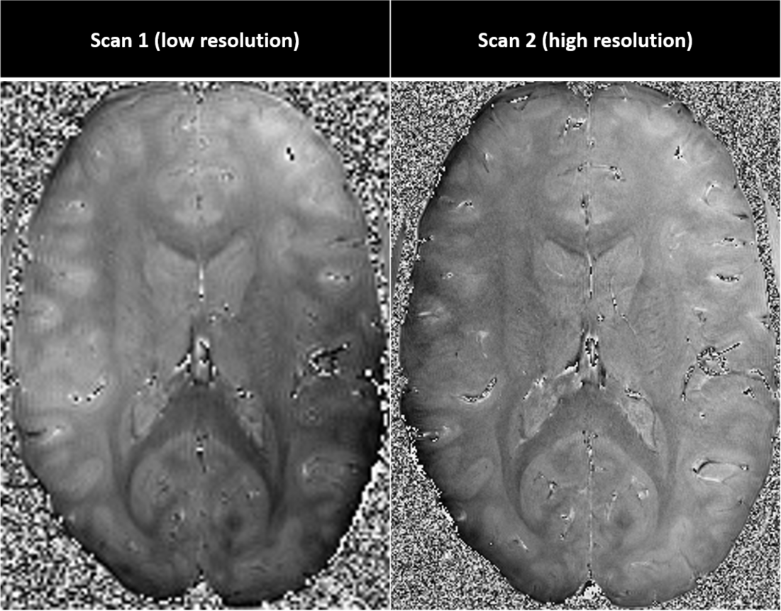
The high resolution Scan 2 has higher noise than Scan 1 despite lower bandwidth due to the substantial reduction in voxel size, but the increased resolution still provides greatly increased detection of small features, which can be seen in Fig. 2.
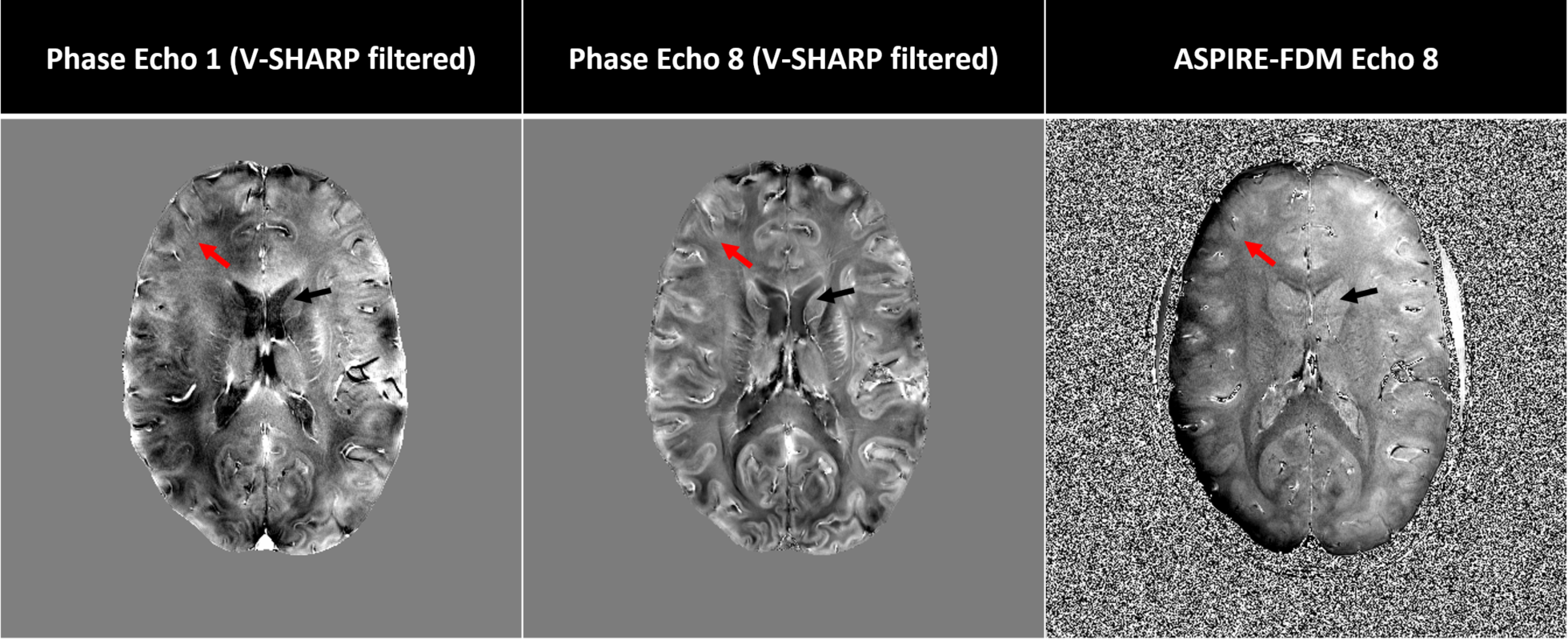
Fig. 3 shows the FDM of the 8th echo, which is calculated from the first and 8th phase image shown. The contrast in the FDM is different to the contrast in either phase image (V-SHARP filtered for better visibility [4]).
Discussion
ASPIRE-FDM with bipolar acquisition allows high resolution, high CNR whole brain FDMs to be acquired in about 10 min, which creates the possibility to use the method in studies with patients. Due to the sensitivity of FDM to microstructure it potentially allows the detection of changes in the microstructure in demyelinating diseases. White matter appears more homogeneous in FDM than in phase images (Fig. 3) potentially making it more feasible to accurately identify Multiple Sclerosis lesions as cortical, juxta-cortical and sub-cortical.
ASPIRE-FDMs contain a spatially non-linear background variation, which is probably caused by higher order effects from non-ideal k-space acquisition due to eddy currents and gradient timing errors.
The biggest contribution to the noise in ASPIRE-FDM images is the phase noise in the first echo. It will be further investigated, if filtering the first phase before subtraction can further decrease the noise without interfering with the produced FDM values.
Another future goal will be the estimation of parameters for the 3-compartment model of white matter from high resolution bipolar acquisitions generated using ASPIRE-FDM.
Conclusion
ASPIRE-FDM improved noise characteristics compared with the FDM methods presented in the literature [2] and can be applied to acquisitions with bipolar readout. The improvements allow higher resolution, reduced measurement time or decreased echo spacing.Acknowledgements
This study was supported by funds of the Oesterreichische Nationalbank Anniversary Fund, Project Number 16213.References
1. Wharton S and Bowtell R. Fiber orientation-dependent white matter contrast in gradient echo MRI. In Proceedings of the National Academy of Sciences, 2012; 109(45):18559-18564
2. Tendler B and Bowtell R. Using frequency difference mapping to assess white matter microstructure in the human corpus callosum. In Proceedings of the 25th Annual Meeting of the ISMRM, 2016. #2849
3. Eckstein K, Dymerska B, Bachrata B, Bogner W, Poljanc K, Trattnig S and Robinson SD. Computationally Efficient Combination of Multi-channel Phase Data From Multi-echo Acquisitions (ASPIRE), Magnetic Resonance in Medicine, 2017; doi: 10.1002/mrm.26963.
4. Li W, Wu B and Liu C. Quantitative susceptibility mapping of human brain reflects spatial variation in tissue composition. Neuroimage, 2011;55(4):1645-1656
Figures