4942
Evaluating Microstructural Integrity of Cortical and Deep Gray Matter over a Season of High School Football Using Diffusion Kurtosis Imaging and Quantitative Susceptibility Mapping1University of California Berkeley, Berkeley, CA, United States, 2Duke University School of Medicine, Durham, NC, United States, 3University of Oklahoma, Norman, OK, United States, 4University of North Carolina at Chapel Hill School of Medicine, Chapel Hill, NC, United States, 5University of Arizona, Tucson, AZ, United States
Synopsis
Over the course of a single high school football season, QSM showed no evidence of microhemorrhage or iron-related changes; however, we observed significant microstructural alterations, as reflected by DKI metrics, in cortical and deep gray matter over the course of a single high school football season. These microstructural changes correlated with the frequency of head impacts, suggesting that DKI imaging of cortical and deep gray matter may yield valuable biomarkers for tracking subclinical traumatic brain injury in contact-sport athletes.
Introduction:
Current estimates indicate that more than 5 million youth and high school athletes are participating in organized American football in the United States [1], most of whom are exposed to repeated head impacts. Subconcussive head impact has also been linked to an increased possibility of long-term cognitive dysfunction and other neurological symptoms [2]. However, potential cerebral microstructural and molecular abnormalities associated with repetitive subconcussive head impacts are not entirely understood. diffusion kurtosis imaging (DKI) and quantitative susceptibility mapping (QSM) have been previously demonstrated promising in probing microstructural and molecular changes in Alzheimer’s disease [3, 4] and aging [5-8]. The first objective of this study was to utilize DKI and QSM for probing microstructural and molecular changes in deep and cortical gray matter in a cohort of high school football players over a single season. By using DKI and QSM at both pre- and post-season, we measured non-Gaussian diffusion and susceptibility changes in the absence of clinically diagnosed concussions. A secondary objective of the study was to examine if the observed changes were associated with subconcussive head impacts sustained during the season.Materials and Methods:
A cohort of seventeen players underwent DKI and QSM scans pre- and post-season. Head impact data was recorded using the Head Impact Telemetry System. DKI and QSM parametric changes in deep and cortical gray matter were examined using FreeSurfer. Association between imaging parametric changes and frequency of frontal head impact was assessed using generalized linear models.Results and Discussion:
In deep gray matter, statistically significant decreases in mean kurtosis (MK) and increases in mean diffusivity (MD) over the season were observed in the thalamus and putamen. In cortical gray matter, decreases in MK were observed in the pars triangularis, inferior parietal, paracentral, and rostral middle frontal cortices. Increases in MD were also observed in the rostral middle frontal cortices. These findings suggested that cumulative head impacts in a single season, even in the absence of a clinically evident concussion, could produce subconcussive microstructural injuries in cortical and subcortical regions. Given the timing of the MRI scans, at the beginning and end of the season, it is possible that much of the microstructural changes observed in the present study, both in cortical and deep gray matter, were in the subacute and chronic recovery phases following repetitive head injury. This may reflect the integrity of neuronal tissue at a time when, presumably, initial inflammatory reactions have subsided and injured axons are in the process of degeneration, which further results in decreased microstructural complexity and increased extracellular diffusion space between neurons.
Magnetic susceptibility values exhibited no significant difference or correlation, suggesting these diffusion changes common within the group were not associated with microhemorrhage- or iron-related injuries. Supporting our finding, a previous study using a mouse model subjected to sustained, cortical impacts also reported no significant change in white matter susceptibility in either the acute or subacute stages of TBI, which suggested that this unchanged susceptibility may reflect a lack of damage to the myelin sheath [9]. Another study reported significant changes in intensities in T2 FLAIR images in military TBI patients without significant changes in susceptibility values, suggesting that the white matter hyperintensities on T2 may not be associated with iron deposition or demyelination [10].
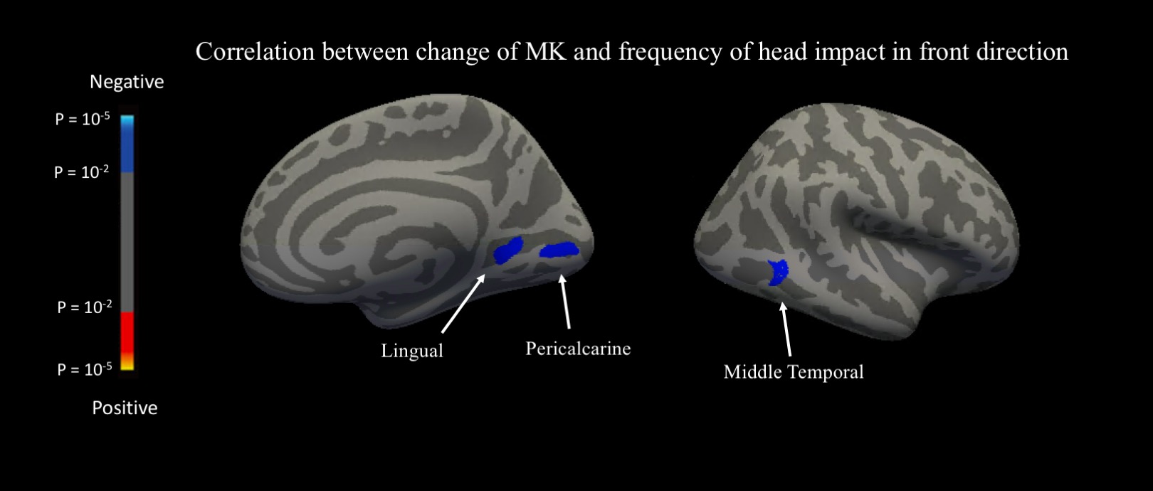
Correlations between change in DKI metric and frequency of frontal impacts were observed in the putamen, caudate, the pericalcarine, lingual and middle temporal cortices. These observations suggested possible vulnerability of the caudate and putamen to head impact in the front direction, which needs to be verified in future studies. Our findings suggested that injury to the opposite side of cranial impact, namely contrecoup injury, could be the leading contributor to an overall decrease of MK. Of note, contrecoup injuries caused by linear accelerations in the anterior–posterior direction have been previously reported in other contact sports such as boxing [11].
Conclusion:
Over the course of a single high school football season, QSM showed no evidence of microhemorrhage or iron-related changes; however, we observed significant microstructural alterations, as reflected by DKI metrics, in cortical and deep gray matter over the course of a single high school football season. These microstructural changes correlated with the frequency of head impacts, suggesting that DKI imaging of cortical and deep gray matter may yield valuable biomarkers for tracking subclinical traumatic brain injury in contact-sport athletes.Acknowledgements
No acknowledgement found.References
1. Daniel, R.W., S. Rowson, and S.M. Duma, Head Impact Exposure in Youth Football. Annals of Biomedical Engineering, 2012. 40(4): p. 976-981.
2. Gardner, A., G.L. Iverson, and P. McCrory, Chronic traumatic encephalopathy in sport: a systematic review. Br J Sports Med, 2014. 48(2): p. 84-90.
3. Gong, N.J., et al., Correlations between microstructural alterations and severity of cognitive deficiency in Alzheimer's disease and mild cognitive impairment: a diffusional kurtosis imaging study. Magn Reson Imaging, 2013. 31(5): p. 688-94.
4. Gong, N.J., et al., Differential microstructural and morphological abnormalities in mild cognitive impairment and Alzheimer's disease: Evidence from cortical and deep gray matter. Hum Brain Mapp, 2017. 38(5): p. 2495-2508.
5. Gong, N.J., et al., Hemisphere, gender and age-related effects on iron deposition in deep gray matter revealed by quantitative susceptibility mapping. Nmr in Biomedicine, 2015. 28(10): p. 1267-1274.
6. Li, W., et al., Differential developmental trajectories of magnetic susceptibility in human brain gray and white matter over the lifespan. Hum Brain Mapp, 2014. 35(6): p. 2698-713.
7. Gong, N.J., et al., Aging in deep gray matter and white matter revealed by diffusional kurtosis imaging. Neurobiol Aging, 2014. 35(10): p. 2203-16.
8. Coutu, J.P., et al., Non-Gaussian water diffusion in aging white matter. Neurobiol Aging, 2014. 35(6): p. 1412-21.
9. Li, W., et al., Spatiotemporal changes in diffusion, T2 and susceptibility of white matter following mild traumatic brain injury. NMR Biomed, 2016. 29(7): p. 896-903.
10. Liu, W., et al. Quantitative Susceptibility Mapping of White Matter Hyperintensities in Patients with Traumatic Brain Injury. in Proc. Intl. Soc. Mag. Reson. Med. 21 (2013)
11. Courville, C.B., Coup-contrecoup mechanism of craniocerebral injuries - Some observations. Archives of Surgery, 1942. 45(1): p. 19-43.
Figures