4941
Characterizing acute, subacute and chronic changes in the brain following sport-related concussion using Diffusion Kurtosis Tensor Imaging1Neurosurgery, Medical College of Wisconsin, Milwaukee, WI, United States, 2Center for Imaging Research, Medical College of Wisconsin, Milwaukee, WI, United States, 3Biophysics, Medical College of Wisconsin, Milwaukee, WI, United States, 4Neurology, Medical College of Wisconsin, Milwaukee, WI, United States
Synopsis
The aim of this study was to characterize acute, subacute and chronic changes in the brain following sport-related concussion in a group of young athletes. Diffusion kurtosis tensor parameters were compared between concussed athletes and controls. The concussed group demonstrated widespread increase in axial kurtosis compared to the controls at the acute time point, which gradually diminished over the course of 14 days. Conversely, alterations in radial kurtosis were mostly in the frontal white and gray matter and occipital gray matter regions. These findings may have important implications for the clinical management of sport-related concussions.
Introduction
Sport-related concussion (SRC) results in rapid onset of specific physical changes and cognitive deficits2,7. However, no abnormalities are seen using conventional MRI. In the absence of established biomarkers, detailed symptom assessment schemes such as SCAT33 have become the gold standard to assess the severity of injury and monitor recovery. Based on such assessments, symptoms typically resolve within 7-10 days of injury. Therefore, SRC has historically been viewed as a minor injury without long-term consequences. However, recent studies suggest that the physiological changes can persist even after symptoms have subsided6. Furthermore, the duration of the physiological recovery is unclear. A better understanding of the trajectory of full recovery is critical for informing symptom management, return-to-play decisions, and prevention of long-term consequences.
To date, most imaging research exploring biomarkers have focused on the brain white matter, since axons are especially vulnerable to shear-strain injury caused by SRC1,4. However, the cortical and subcortical gray matter could also be affected by such impacts. Therefore, we studied changes in the whole brain in injured athletes using diffusion kurtosis tensor imaging (DKTI).
Methods
60 concussed high school and college football players (mean age=17.9; SD =1.73) and 60 contact-sport controls (mean age=18.2; SD=1.61) participated in this study. The study was approved by the IRB and written consents were obtained from subjects. The participants underwent MRI scans and were also administered SCAT3 and Standardized Assessment of Concussion and Balance Error Scoring System within 48 hours and then followed up at 8 days, 14 days and 45 days after injury. Data were acquired using GE MR750 3T MRI scanner. DKTI was collected as part of multimodal imaging protocol using a single-shot SE-EPI sequence with 3mm isotropic voxels, four non-diffusion weighted images and diffusion-weighted images with b=1000s/mm2 and 2000s/mm2 (30 diffusion directions for each shell). Then, FA, MD, Dax, Drad (axial and radial diffusivity), MK, Kax and Krad (mean, axial and radial kurtosis) maps were calculated. White matter differences between groups at each time point were studied using TBSS workflow8. Cortical and subcortical gray matter differences were analyzed using a voxel-based analysis approach. For cortical gray matter analysis, each DKTI image was registered to the standard template and masked with a binary mask generated from Harvard-Oxford cortical atlas. Subcortical gray matter differences were analyzed using the same approach using Harvard-Oxford subcortical gray matter atlas. Gray matter images were smoothed with 4mm Gaussian for voxel-based analysis. All statistic maps were thresholded at p<0.05, corrected for multiple comparisons. Test-retest reliability of longitudinal data was assessed using concordance correlation coefficient (CCC)5 using repeat scans from control subjects. Average CCC was 0.92 for MD, 0.88 for Krad and 0.82 for Kax.Results
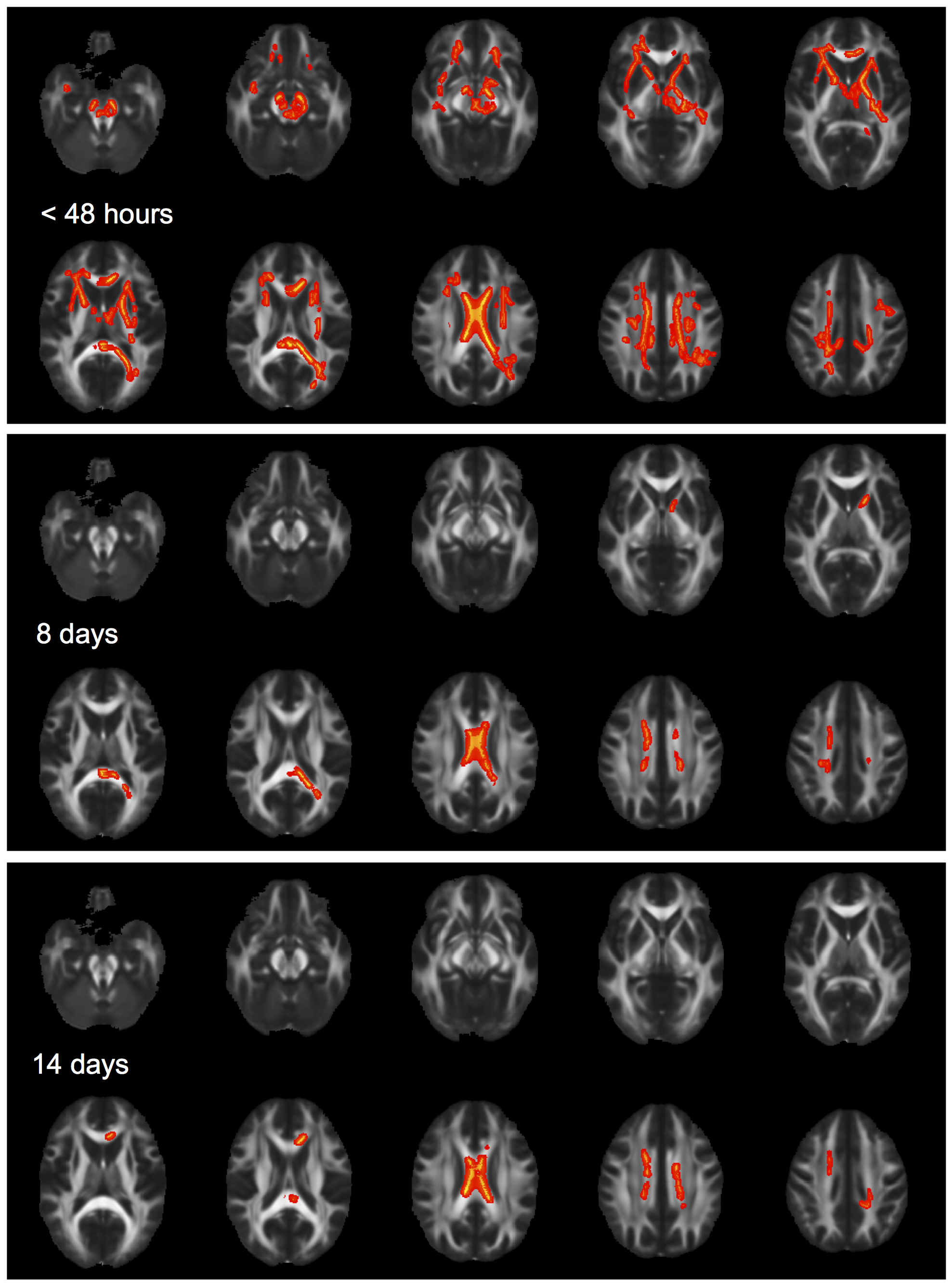
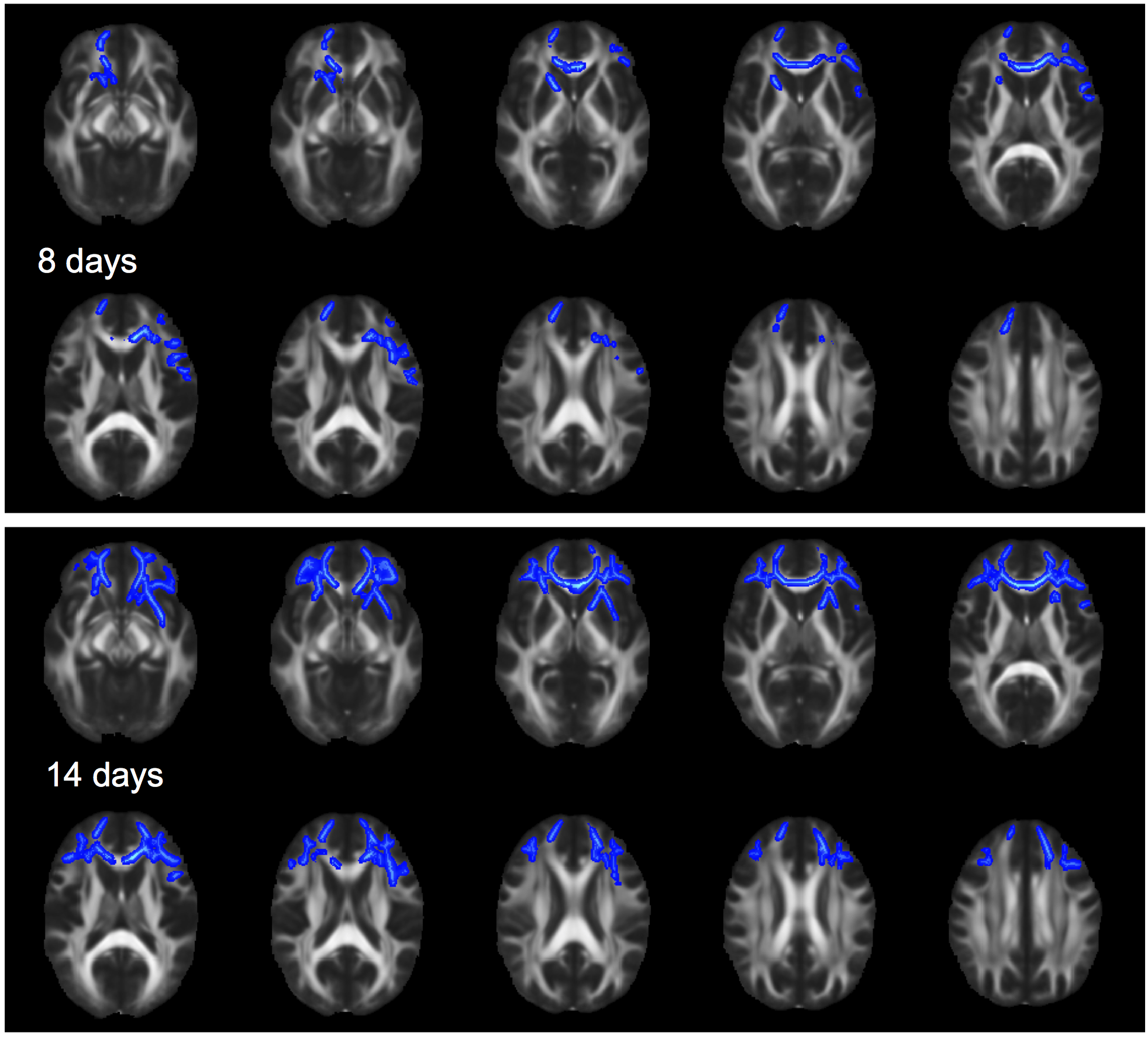
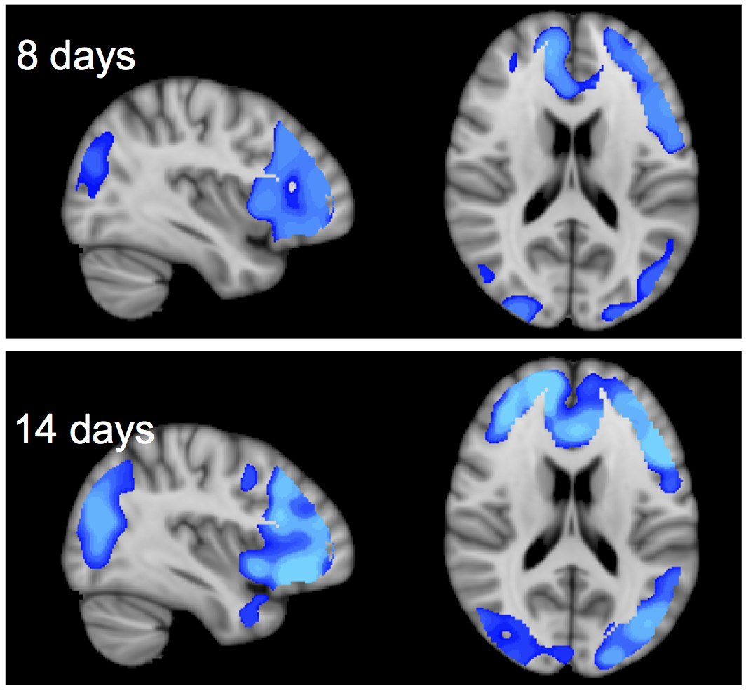
Concussed athletes had significantly higher Kax in major white matter fiber tracts compared to controls (Fig.1). The effects were widespread at the acute time point and gradually receded to the corpus callosum by day 14 and diminished by day 45. On the other hand, Krad showed a delayed response, both in white matter and cortical gray matter. No differences were seen at the acute time point; but concussed subjects showed decreased Krad mostly in frontal white matter and gray matter and occipital gray matter on days 8 and 14, which diminished by day 45 (Fig.2 and 3). Other DKTI metrics did not show statistically significant differences between the two groups at any time point. Furthermore, no differences were detected in subcortical gray matter.Discussion and conclusion
These findings indicate that the concussed athletes have significant white matter and gray matter alterations during the first 14 days after injury, despite normalization of clinical symptoms within a week after injury. Kax and Krad showed different trajectories of progression during the 14-day window, which might reflect region-dependent injury mechanisms. Interestingly, Krad changes were mainly confined to the gray matter in the occipital areas and concurrent white matter changes were not seen here.
It should be noted that the voxel-based analysis used spatially smoothed images and a probabilistic gray matter mask, which were necessary to account for variations in gyral and sulcal anatomy across subjects. Thus, peripheral white matter was partially included inevitably in the analyses. This is not necessarily a shortfall, as peripheral white matter is also vulnerable to such injuries. Our measurements reveal an average injury effect in the cortical gray matter, including peripheral axonal branches connecting to those cortical regions.
These findings have implications for determination of recovery following SRC and concussion management. Further studies are needed to investigate the long-term impact of these white matter changes.
Acknowledgements
This project was supported by the National Center for Advancing Translational Sciences, National Institutes of Health (8UL1TR000055 and 1UL1-RR031973-01), the US Army Medical Research and Materiel Command (W81XWH-12-1-0004), and the NFL-GE Head Health Challenge.References
1. Gentry, L. R. (1994) Imaging of closed head injury. Radiology 191, 1-17.
2. Giza, C. C. and Hovda, D. A. (2014) The new neurometabolic cascade of concussion. Neurosurgery 75 Suppl 4, S24-33.
3. Guskiewicz, K. M., Register-Mihalik, J., McCrory, P., McCrea, M., Johnston, K., Makdissi, M., Dvorák, J., Davis, G. and Meeuwisse, W. (2013) Evidence-based approach to revising the SCAT2: introducing the SCAT3. Br J Sports Med 47, 289-293.
4. Holbourn, A. H. S. (1943) Mechanics of head injuries. The Lancet 242, 438-441.
5. Lin, L. I. (1989) A concordance correlation coefficient to evaluate reproducibility. Biometrics 45, 255-268.
6. McCrea, M., Prichep, L., Powell, M. R., Chabot, R. and Barr, W. B. (2010) Acute effects and recovery after sport-related concussion: a neurocognitive and quantitative brain electrical activity study. J Head Trauma Rehabil 25, 283-292.
7. McCrory, P., Meeuwisse, W. H., Aubry, M., Cantu, B., Dvorák, J., Echemendia, R. J., Engebretsen, L., Johnston, K., Kutcher, J. S., Raftery, M., Sills, A., Benson, B. W., Davis, G. A., Ellenbogen, R. G., Guskiewicz, K., Herring, S. A., Iverson, G. L., Jordan, B. D., Kissick, J., McCrea, M., McIntosh, A. S., Maddocks, D., Makdissi, M., Purcell, L., Putukian, M., Schneider, K., Tator, C. H. and Turner, M. (2013) Consensus statement on concussion in sport: the 4th International Conference on Concussion in Sport held in Zurich, November 2012. Br J Sports Med 47, 250-258.
8. Smith, S. M., Jenkinson, M., Johansen-Berg, H., Rueckert, D., Nichols, T. E., Mackay, C. E., Watkins, K. E., Ciccarelli, O., Cader, M. Z. and Matthews, P. M. (2006) Tract-based spatial statistics: voxelwise analysis of multi-subject diffusion data. Neuroimage 31, 1487-1505.
Figures