4940
Within-Subject Characterization of Sports Concussion with Serial Diffusion MRI and Spherical Mean Technique1Neurosurgery, Medical College of Wisconsin, Milwaukee, WI, United States, 2Radiology, Medical College of Wisconsin, Milwaukee, WI, United States
Synopsis
Diffusion tensor imaging is a promising biomarker of mild Traumatic Brain Injury and concussion, but it has not been refined sufficiently for diagnosis in individual subjects. In this study, we demonstrate that spherical mean technique (SMT) diffusion MRI can detect post-concussion changes in single subjects over the acute recovery period.
Introduction
Diffusion tensor imaging (DTI) has consistently identified changes in the brain as a result of sports concussion at a group level1-3. However, its clinical significance is still limited by the inability to detect injury within individual subjects4. Variations in individual brain anatomy and the lack of pre-injury baseline MRI makes detection challenging. In this work, we utilize serial MRI post-concussion to implicate the effects of concussion within individual subjects. Additionally, the advantages of spherical mean technique5 (SMT) diffusion MRI to reduce brain anatomical variability were evaluated. The results demonstrate that serial MRI in combination with SMT enables detection of post-concussive changes in individual subjects and reveals interesting spatial and temporal patterns of suspected injury and recovery in the concussed brain.
Methods
All procedures were approved by the Institutional Review Board. Participants were recruited from an ongoing prospective study of recovery following concussion in high school and collegiate athletes, Project Head to Head II, with MRI at 4 timepoints(1, 7, 14, and 45 days) post-concussion(N=39) and identical timepoints in matched control participants(N=41).
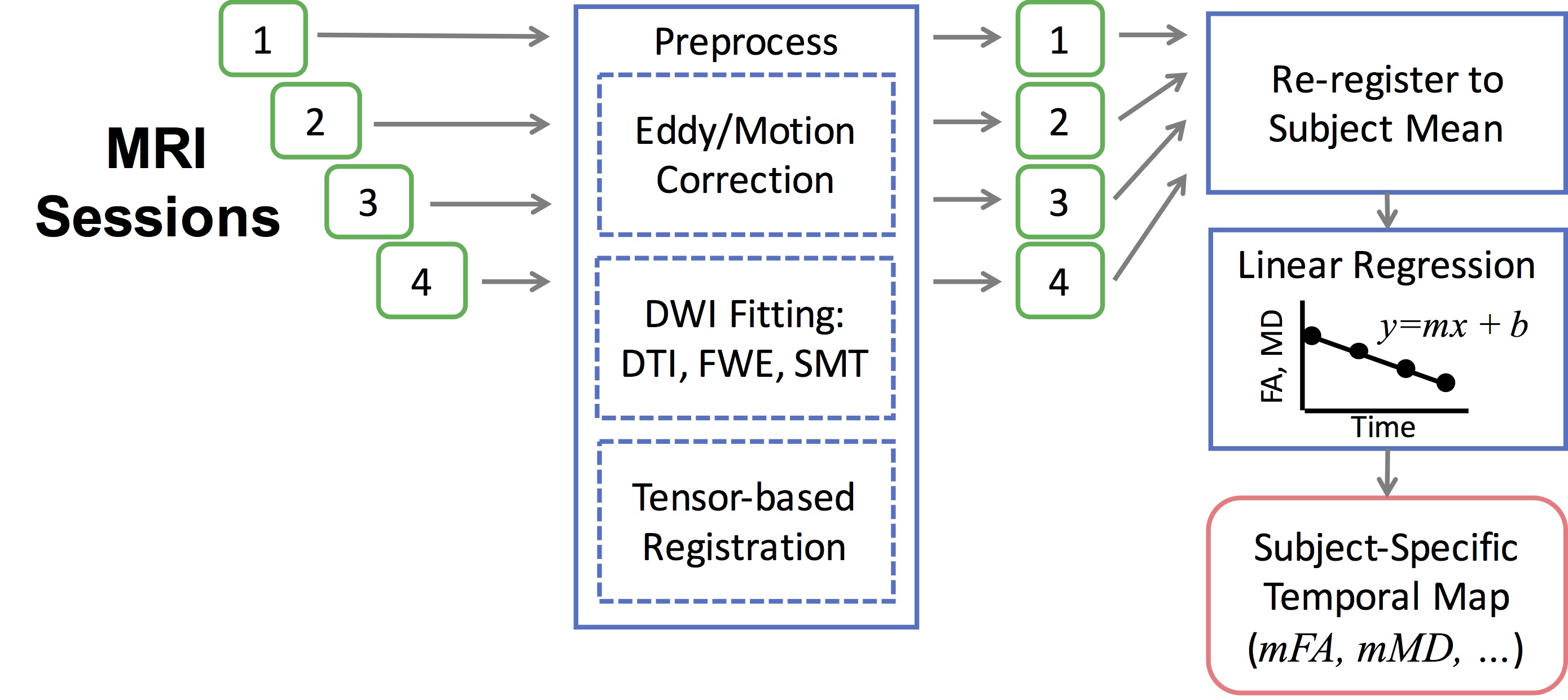
MRI was performed on a 3T GE Discovery MR750 witha 32-channel array coil. Diffusion weighted images were acquired with a single shot echo planar imaging (TR/TE: 5250/67) at 2 b-values (1000 and 2000 s/mm2) along 30 directions. Initial pre-processing(Fig 2) included correction for motion, eddy-current, and susceptibility distortion(FSL5.0.9), followed by DTI(FSL) and SMT(Matlab) estimation, and tensor-based spatial registration(DTI-TK) to MNI space using the IIT DTI template(v4.1)6. For each subject, all 4 timepoints were averaged and each timepoint was re-registered to the subject's mean. Parameter maps (DTI: FA, MD, RD, and AD; SMT: Daxonal, Dextra, v, and FA) were transformed, and for each subject, the temporal change in each voxel (slope: m) was estimated using linear regression over time. Both the raw and absolute values were assessed for statistical significance using a non-parametric groupwise comparison with correction for the false discovery rate (FDR). A subsequent region of interest analysis encompassing the thalamus and internal capsule was assessed for within-timepoint group differences in raw values and z-scores.
Results
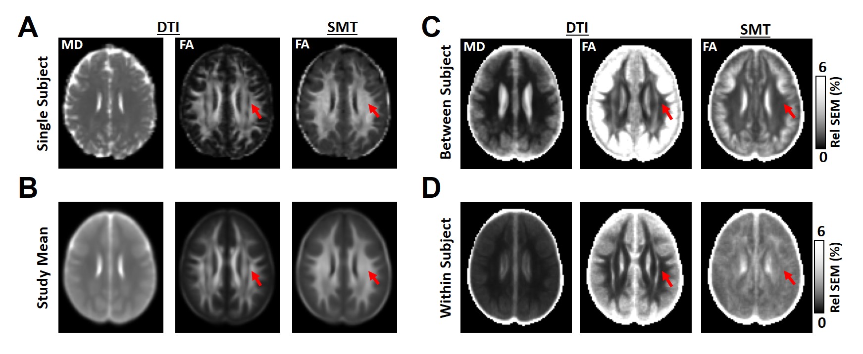
As expected (Fig. 1), maps of FASMT exhibit minimal "crossing-fiber" regions in the brain compared to FADTI. Across all control subjects, the within-subjects relative standard error of the mean (RelSEM) across the 4 timepoints was lower for all metrics than the between-subjects RelSEM, with the greatest reductions noted in gray matter. Although the within-subject FASMT variability was greater than FADTI, it was nearly uniform in the brain whereas FADTI had clear regional differences in variance.
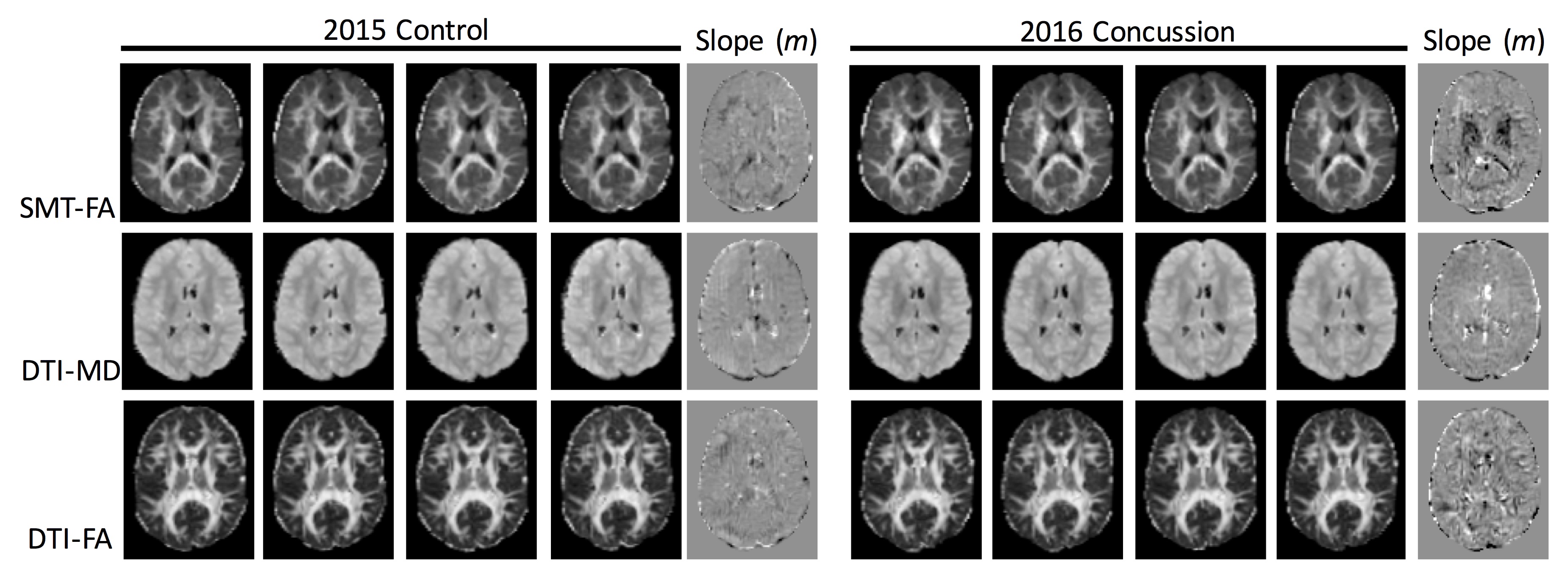
In a single example subject (Fig. 3), maps of the slope over the initial 45 day post-concussive period revealed regions of high change in FASMT primarily in deep brain structures. The changes in MDDTI were less pronounced, and FADTI did not reveal any consistent qualitative patterns. Qualitative comparisons between the same subject as a prior control demonstrate the greater sensitivity to changes post-concussion.
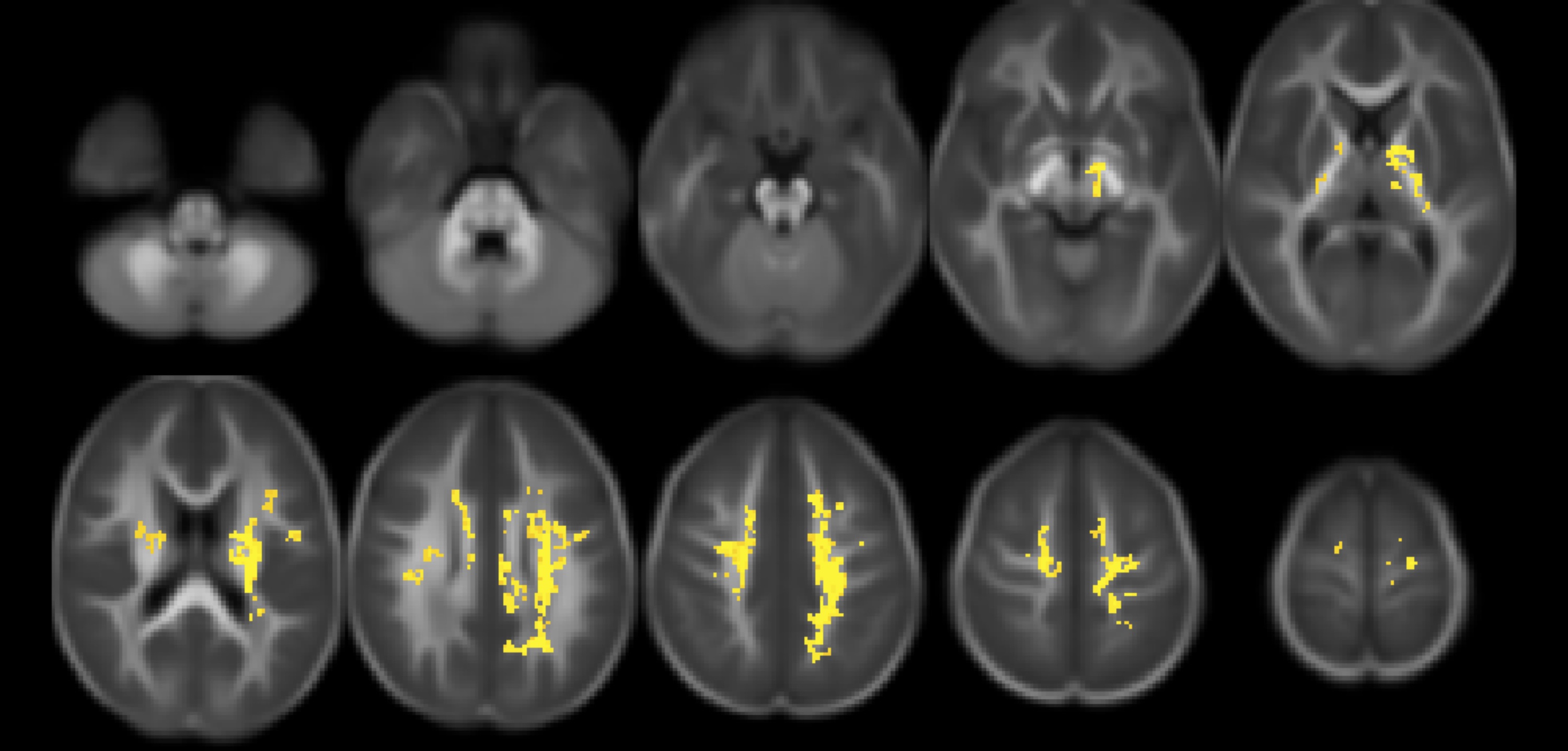
In the group analysis, there was no significant differences in the slope over time in any of the metrics the concussed group compared to the control group (not shown). However, there was a significant change in the absolute change (|m|) in FASMT (Fig. 4) within the central and deep brain structures. Changes were also evident (not shown) in v, Daxonal, and Dextra.
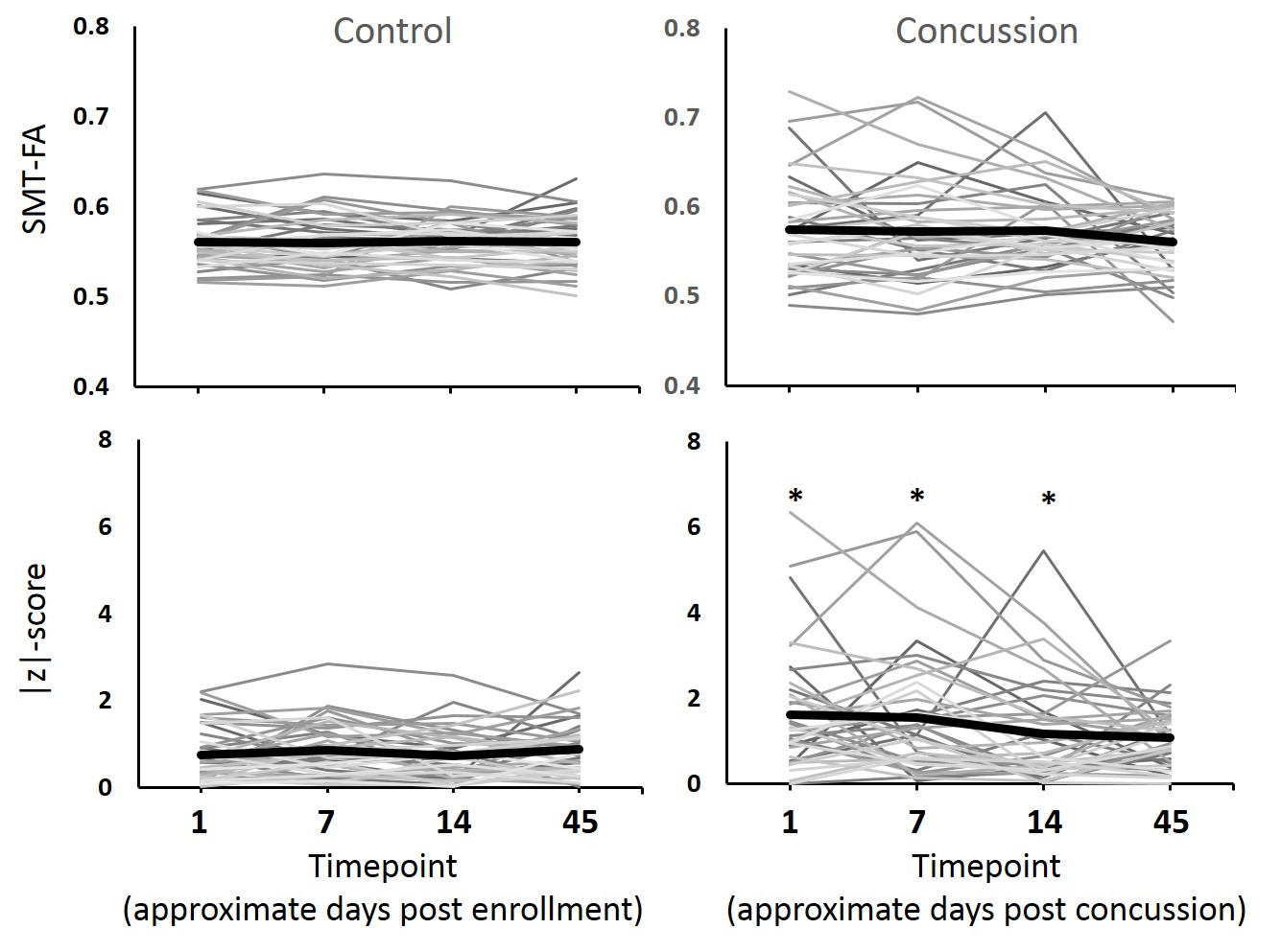
A subsequent region of interest analysis in the thalamic and internal capsule region (Fig. 5) revealed that while the control subjects had stable timecourses in FASMT, the concussed subjects revealed striking variability over time. While both decreases and increases in FASMT were evident within individual subjects, the directionality was consistent within each subject. These changes tended to revert to the mean over time, as significant differences in the absolute z-scores were evident at 1 (p=0.0004), 7 (p=0.006), 14 (p=0.035), but not 45 days post-injury (p=0.16). These corresponded to 11 (28%), 10 (26%), 8 (21%), and 3 (8%) of 39 concussed subjects greater than 2 standard deviations from the control values at each timepoint compared to 3, 1, 1, and 2 of 41 control subjects at the same timepoints.
Discussion
These results demonstrate several interesting findings: 1)SMT reduces the spatial variability within the brain; 2)Within-subject longitudinal analysis has reduced variability compared to between-subject analysis; 3)FASMT reveals evidence of single-subject and significant group differences associated with concussion; 4)Both increased and decreased FA are evident after concussion5, but appear to be consistent within subjects and tend to normalize over time.
Collectively, this study demonstrates that concussion elicits changes in deep brain structures that can be detected in individual subjects that recover over time. The significance of the brain regions involved, directionality of the changes, and relationships to clinical impairment and recovery remain to be elucidated.
Acknowledgements
This work was supported by the U.S. Army Medical Research and Materiel Command under award number W81XWH-12-1-0004. Opinions, interpretations, conclusions, and recommendations are those of the authors and are not necessarily endorsed by the U.S. Army.References
1Mustafi, SM, et al. J Neurotrauma. 2017. 2Meier TB, et al. Hum Brain Mapp. 2016. 3Lancaster MA, et al Hum Brain Mapp. 2016. 4Amyot F, et al. J Neurotrauma. 2015. 5Kaden E, et al. Magn Reson Med. 2016. 6Varentsova A, et al. Neuroimage. 2014. 7Ling, JM, et al. Brain. 2012.
Figures