4911
Simultaneous multislice spiral cine DENSE MRI1Biomedical Engineering, University of Virginia, Charlottesville, VA, United States, 2Medicine, University of Virginia, Charlottesville, VA, United States, 3Radiology, University of Virginia, Charlottesville, VA, United States
Synopsis
Spiral cine DENSE is an established method for imaging myocardial strain, however a relatively long acquisition time is a limitation. We developed a simultaneous multislice (SMS) method to accelerate spiral cine DENSE imaging. For SMS excitation we employed CAIPIRINHA phase modulation of the multiple slices. For the SMS reconstruction, we implemented a modified iterative conjugate gradient sensitivity encoding (CG-SENSE) method. Simulations and experiments in phantoms show that a 10-15% error occurs when imaging two slices simultaneously. Two-slice volunteer SMS images showed close agreement with separately-acquired single-slice images. SMS with CG-SENSE may be an effective means to accelerate spiral cine DENSE.
Purpose
Spiral cine displacement encoding with stimulated echoes (DENSE) is an established method for imaging myocardial strain [1]. The advantages of spiral cine DENSE are rapid strain analysis and higher spatial resolution compared with myocardial tagging; however, a longer acquisition time is a limitation [2]. Simultaneous multislice (SMS) imaging has made a major impact in MRI of the brain. However, to date SMS has not seen widespread application in cardiac MRI and there has not been extensive effort devoted to reconstruction of SMS spiral data [3]. We sought to develop an SMS spiral cine DENSE method in order to accelerate DENSE imaging.Methods
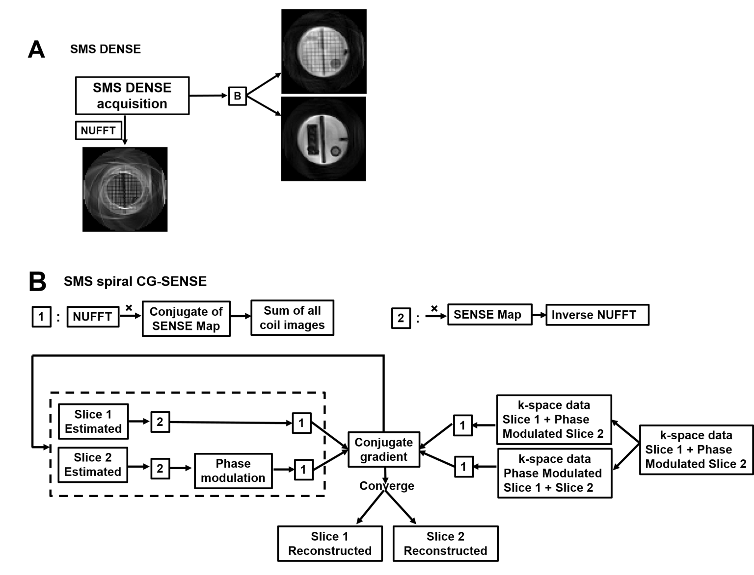
For SMS DENSE RF excitation we employed CAIPIRINHA phase modulation of the multiple slices [3]. For the spiral SMS reconstruction (Fig. 1), we implemented a modified iterative conjugate gradient sensitivity encoding (CG-SENSE) method based on a method previously employed for radial CAIPIRINHA [4]. This method requires sensitivity maps for each slice, and these were acquired using separate breathold acquisitions for each slice. We used phantoms to evaluate these methods, including simulations of SMS spiral DENSE using separately acquired slices, as well as true SMS acquisitions where slices were acquired simultaneously. We also evaluated the SMS spiral cine DENSE method in a human volunteer, where the SMS images were compared to separately-acquired DENSE images at matched slice locations. For phantom and human scans, RF coils had 30-34 channels.Results
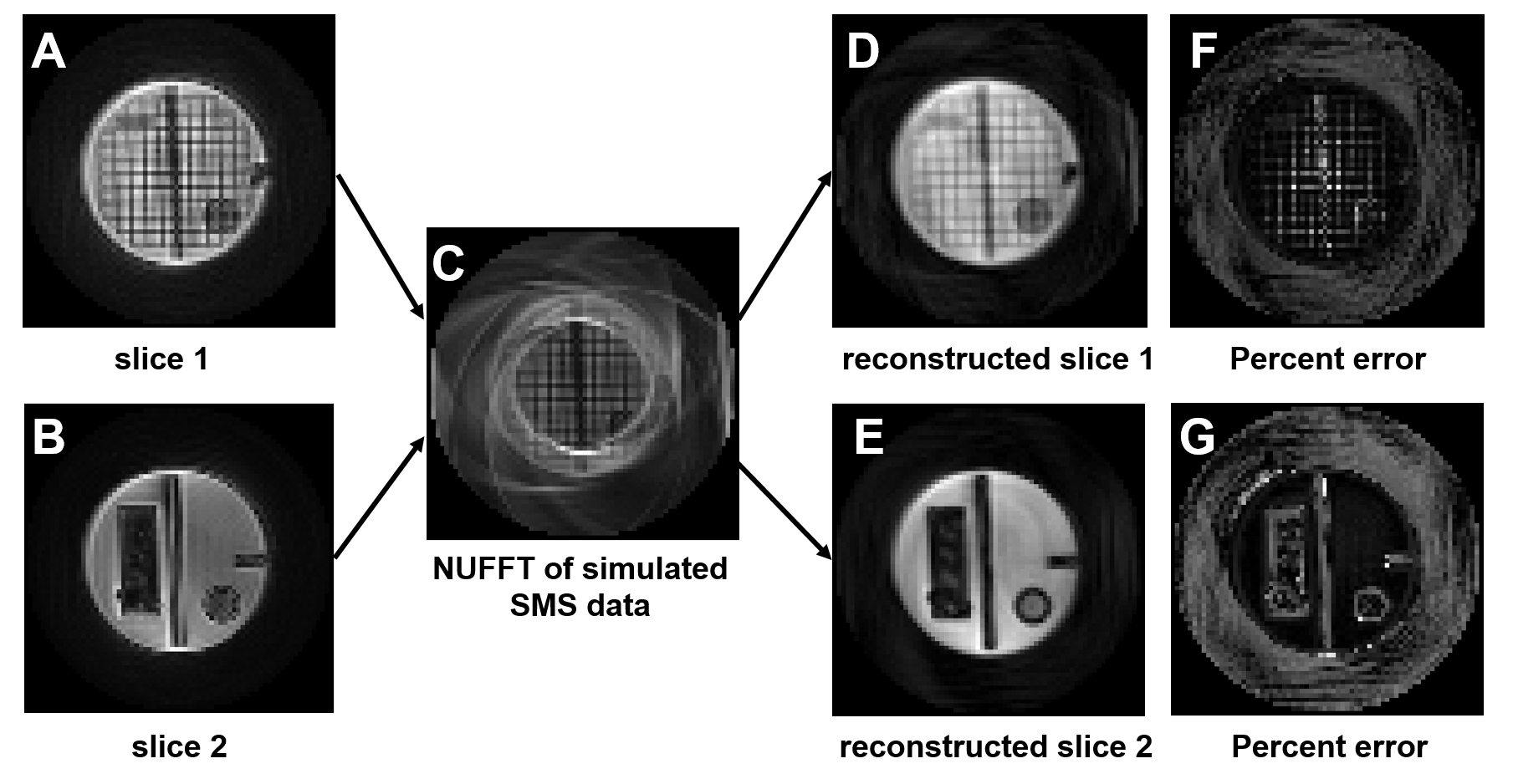
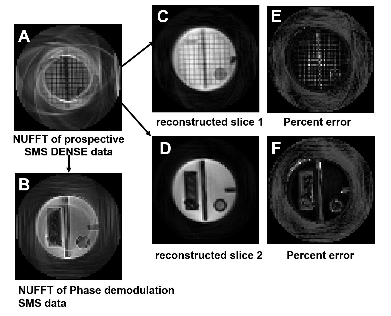
Figure 2 shows the results of simulations of SMS spiral DENSE using two separately acquired slices (Fig. 2A,B). Fig. 2C shows the simulated SMS image reconstructed using NUFFT [5]. Fig. 2D,E show the two images of different slices recovered after application of the spiral CG-SENSE algorithm, and Figure 2F,G shows the difference between the recovered images and the original images. The average percent errors in signal intensity in homogeneous regions were 13.66% and 11.62% for the two slices, respectively. The evaluation of two simultaneously acquired phantom slices is shown in Figure 3. Specifically, Figure 3A,B show the NUFFT and phase-demodulated NUFFT of the SMS raw data, depicting initial estimates of the two separate slices. Figure 3C,D shows the two images of different slices recovered after application of the spiral CG-SENSE algorithm, and Figure 3E,F shows the difference between the recovered images and the separately acquired images shown in Figure 2A, B. The average percent errors in homogeneous regions were 12.03% and 9.94% for the two slices, respectively.
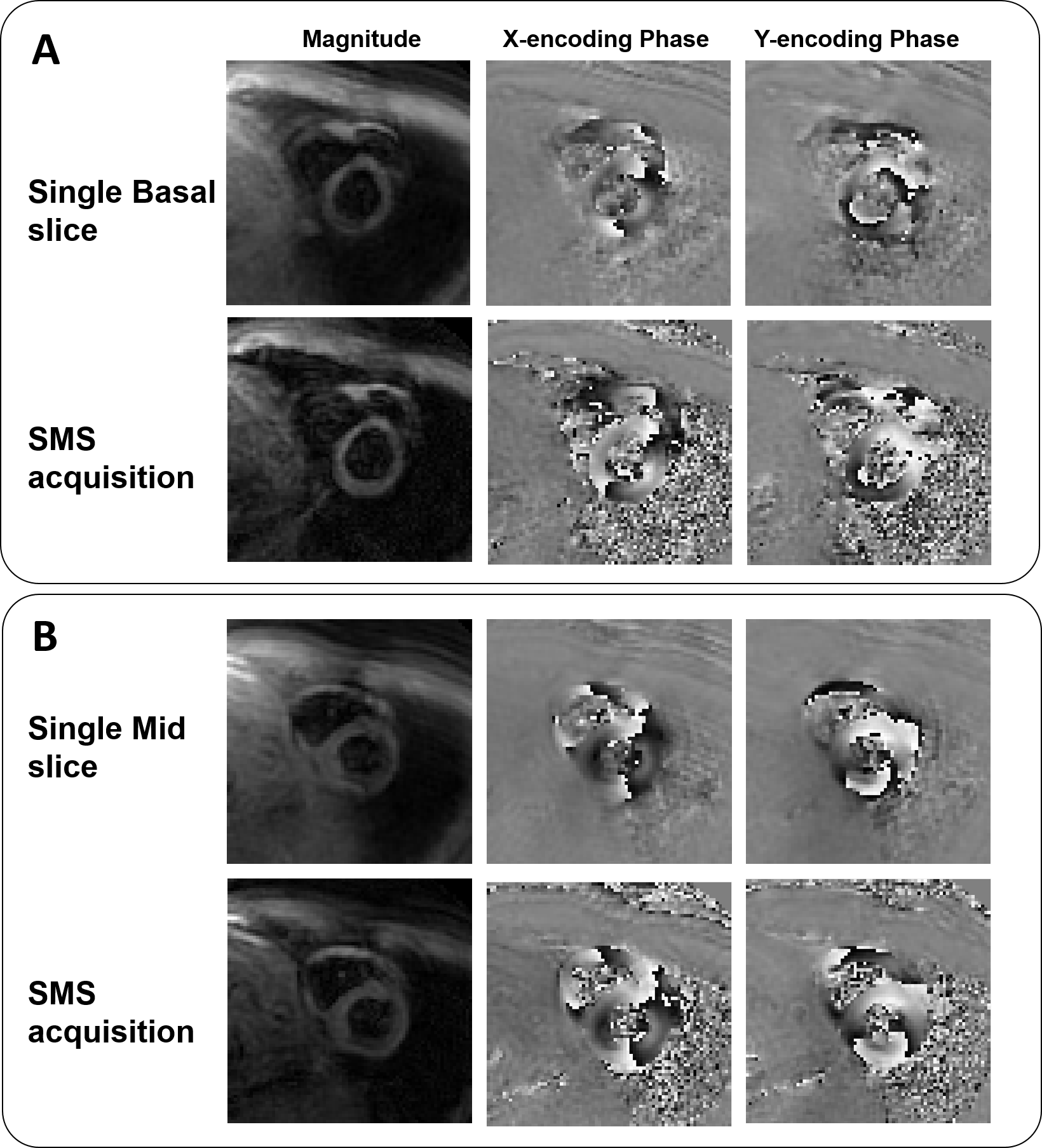
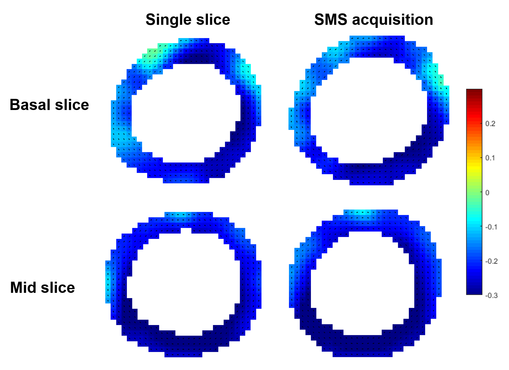
Results from a human volunteer are shown in Figure 4. Specifically, for a reference, separately-acquired spiral cine DENSE short-axis images at basal and mid-ventricular locations are shown in Figure 4A and Figure 4B, and CG-SENSE-recovered SMS spiral DENSE images at the same location are shown in Figure 4A,B. Qualitatively, there is good agreement between the SMS spiral DENSE images and the separately-acquired spiral cine DENSE images. Similarly, myocardial strain maps in Figure 5 show a close correspondence between the SMS and separately acquired slices.
Discussion
SMS with CG-SENSE may be an effective means to accelerate spiral cine DENSE imaging. Phantom data suggest that errors may be on the order of only 10-15% when acquiring two slices simultaneously. Volunteer imaging showed promising initial results. Ongoing work includes acquiring and recovering more than two slices simultaneously, autocalibration methods for sensitivity maps, modifying the reconstruction to include a constraint related to temporal sparsity in the cardiac phases dimension, and evaluating SMS spiral DENSE more thoroughly in phantoms and volunteers.Acknowledgements
Research support from Siemens Healthineers.References
1. Zhong et al. MRM. 2010;64(4):1089-1097. 2. Chen et al. JCMR, 2016; 18(1):38-51. 3. Yang et al. ISMRM, 2017; #3242. 4. Yutzy et al. MRM, 2011; 65(6): 1630-1637. 5. Fessler et al. IEEE T Signal Proces, 2003; 51(2): 560-574.Figures