4886
Contrast steady state myocardial scar imaging in a chronic porcine infarct model1Division of Imaging Sciences and Biomedical Engineering, King's College London, London, United Kingdom, 2Siemen's Healthcare, Erlangen, Germany
Synopsis
Contrast steady state (CSS) may be achieved using continuous contrast infusion following bolus. This technique was used to acquire high resolution late gadolinium enhanced (LGE) scar imaging of 8 pigs with chronic myocardial infarction. CSS imaging allowed extended acquisition to facilitate high-resolution 3D LGE imaging with improved signal-to-noise, contrast-to-noise and overall image quality. CSS may also be used in experimental studies, when total gadolinium bolus is not restricted, to directly compare imaging sequences acquired using consistent contrast distributions.
Introduction
High resolution 3D cardiac magnetic resonance (CMR) scar
imaging is usually acquired following a bolus of gadolinium based contrast
agent and based on the assumption that contrast concentration in blood pool and
myocardial tissue does not change significantly during the CMR acquisition.
This limits the duration over which imaging can be acquired and impacts the
resolution of imaging that can be achieved. The use of an infusion to achieve contrast
equilibrium has been previously reported in the calculation of extracellular
volume (ECV) maps(1). We hypothesised that continuous
contrast infusion could be used during late gadolinium enhancement (LGE)
imaging to establish a stable myocardial T1, thereby allowing
extended acquisition for high-resolution 3D fibrosis imaging without
significant change in inversion times (TI) for blood and myocardium. Methods
All imaging was performed at 1.5T (MAGNETOM Aera, Siemens
Healthcare, Germany). Eight pigs with chronic myocardial infarction were
studied. Ten minutes after 0.1mmol/kg gadobutrol (Gd) bolus, TI scout was
acquired in a single short axis slice. Clinical standard 2D-LGE scar sequences
were acquired in 8 short axis slices using an inversion recovery (IR) sequence with
phase-sensitive IR (PSIR) reconstruction (SSFP; linear k-space reordering;
TE/TR/α:
1.21ms/3ms/45°; slice thickness: 6mm; in plane resolution: 1.4x1.4mm2).
Fifteen minutes following Gd bolus, 0.0011mmol/kg/min Gd infusion was commenced.
TI-scout was repeated at regular intervals. 30 minutes following Gd bolus,
isotropic navigator-gated ECG-triggered 3D IR sequence was acquired (SSFP; coronal
orientation; linear k-space reordering; TE/TR/α: 1.58ms/3.6ms/90°;
gating window = 7mm; resolution 1.2x1.2x1.2mm3). Scar imaging was
reviewed by 2 experienced observers who graded it on a 4 point scale according
to image sharpness, image contrast, quality of myocardial nulling, complexity
of scar pattern and overall scan quality. Signal-to-noise ratio (SNR) and contrast-to-noise
ratio (CNR) were estimated using mean signal within blood, remote (healthy)
myocardium and scar and standard deviation of signal within lung. Results
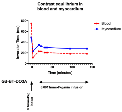
Contrast steady state (CSS, <5% variation in blood/myocardium
TI) was achieved in 7 pigs and maintained for up to 150 minutes (figure 1). Mean
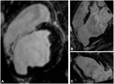
acquisition for CSS 3D-LGE imaging was 51 minutes. CSS 3D-LGE imaging was
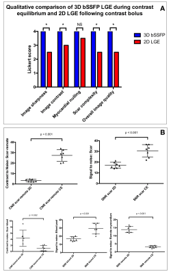
acquired using a total Gd dose of <0.2mmol/kg. SNRScar, SNRBlood
and CNRScar-remote was higher in CSS 3D-LGE imaging than the 2D-LGE
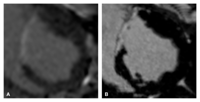
imaging (p<0.001) (figure 3). SNRRemote was lower in the CSS 3D-LGE
than the 2D-LGE imaging (p<0.05), indicating more effective nulling of the
myocardium. There was a significant median increase in sharpness (4.0 vs
2.5, p=0.026), contrast (4.0 vs 3.0, p=0.024), appreciation of scar complexity
(4.0 vs 2.5, p=0.038) and overall scan quality (4.0 vs 2.5, p=0.041) in the CSS
3D-LGE imaging compared to the post Gd bolus 2D imaging (figure 3).Discussion
We have demonstrated that CSS imaging facilitates extended
acquisition times without requiring contrast doses above those used in routine
clinical practice(2). Despite the prolonged
acquisition, SNR in scar, healthy myocardium and blood pool were
significantly above that observed in clinical standard 2D LGE imaging, acquired
in under 5 minutes. Higher SNR and CNR between scar
and healthy tissue will facilitate accurate identification of scar. Higher
signal in the blood pool reduced the contrast between blood pool and scar which
may make accurate identification of the endocardium challenging in areas of
transmural scar. Black blood scar imaging techniques may be helpful to overcome
this issue. The maintenance of CSS for up to 150 minutes demonstrates that this
technique may be used in experimental conditions, where total Gd dose is not
restricted, to directly compare imaging sequences by allowing extended imaging
of the same tissue under consistent contrast conditions. Conclusion
CSS allows extended acquisition to facilitate high-resolution
3D LGE imaging with improved SNR, CNR and overall image quality. CSS-LGE
imaging may be valuable when detailed myocardial substrate assessment is
required, for example when planning cardiac electrophysiology procedures. In
addition, CSS-LGE imaging provides an experimental model in which post-contrast
sequences may be compared with consistent contrast distributions.Acknowledgements
No acknowledgement found.References
1. Flett AS, Hayward MP, Ashworth MT, Hansen MS, Taylor AM, Elliott PM, McGregor C, Moon JC. Equilibrium contrast cardiovascular magnetic resonance for the measurement of diffuse myocardial fibrosis: Preliminary validation in humans. Circulation. 2010;122:138–144.
2. Nacif MS, Arai AA, Lima JA, Bluemke DA. Gadolinium-enhanced cardiovascular magnetic resonance: administered dose in relationship to united states food and drug administration (FDA) guidelines. J Cardiovasc Magn Reson. 2012;14:18.
Figures


