4869
Estimation of late gadolinium enhancement of myocardial infarction with phase sensitive inversion recovery balanced steady state free precession (PSIR-bSSFP)1Radiology, Tokyo Metropolitan Police Hospital, Tokyo, Japan, 2Graduate school of Medicine, Division of Diagnostic Image Analysis, Tohoku University, Miyagi, Japan, 3MR Clinical Science, Philips Japan, Tokyo, Japan, 4Radiology, Kenkoin Clinic, Tokyo, Japan, 5Diagnosis of radiology, Tokyo Metropolitan Police Hospital, Tokyo, Japan
Synopsis
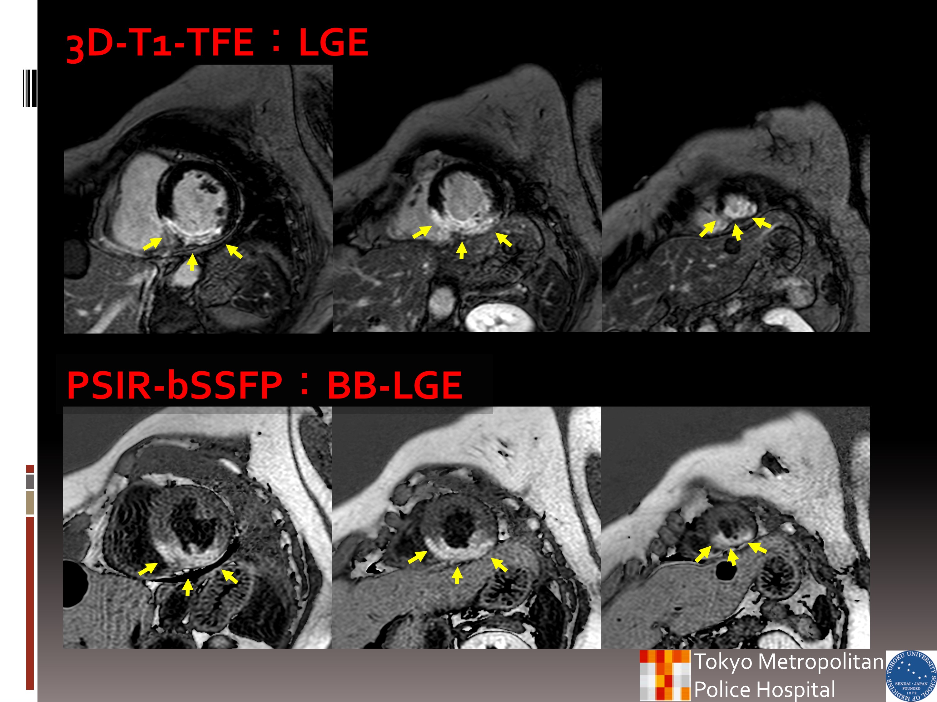
The late gallium enhancement (LGE) image showing an expression amount having a significant difference for improvement wall motion or not is estimated from contrast enhancement area in patients with cardiovascular disease. However, left ventricle blood (LVB) with contrast wash-out can have similar T1 value to scar, and a fabrication sub-endocardial scar is often deluded as LVB. The phase sensitive inversion recovery balanced steady state free precession (PSIR-bSSFP) is possible to obtain black blood and contrast enhancement image. Our study of PSIR-bSSFP was propose BB-LGE that feasible visualize of myocardial hyper-enhancement area with LV blood suppression.
Aims
A T1-weighted turbo field echo (T1-TFE) sequence with phase sensitive inversion recovery (PSIR) provides a wider range of signal intensity than conventional inversion recovery T1-TFE (IR-T1-TFE) sequences by phase correction combining negative and positive longitudinal magnetization in the image. T1-TFE sequence with PSIR is applied to a late gadolinium enhancement (LGE) for scar/fibrosis in patients with cardiovascular disease. However, blood can have similar T1 value to scar by contrast wash-out, and often be depicted as a fabrication sub-endocardial scar. The characteristic of balanced steady-state-free-precession (bSSFP) is the insulation from the influence of blood flow, and the higher contrast of LGE of scar/fibrosis. The bSSFP using PSIR method (PSIR-bSSFP) has been reported to provide a black blood (BB) image that is independent from blood flow velocity, directions, or inversion time (TI). The purpose of this study was to compare black blood (BB)-LGE using a PSIR-bSSFP and the usual LGE using 3D-IR-T1-TFE method for detection of scar extent of myocardial infarction.Methods and materials
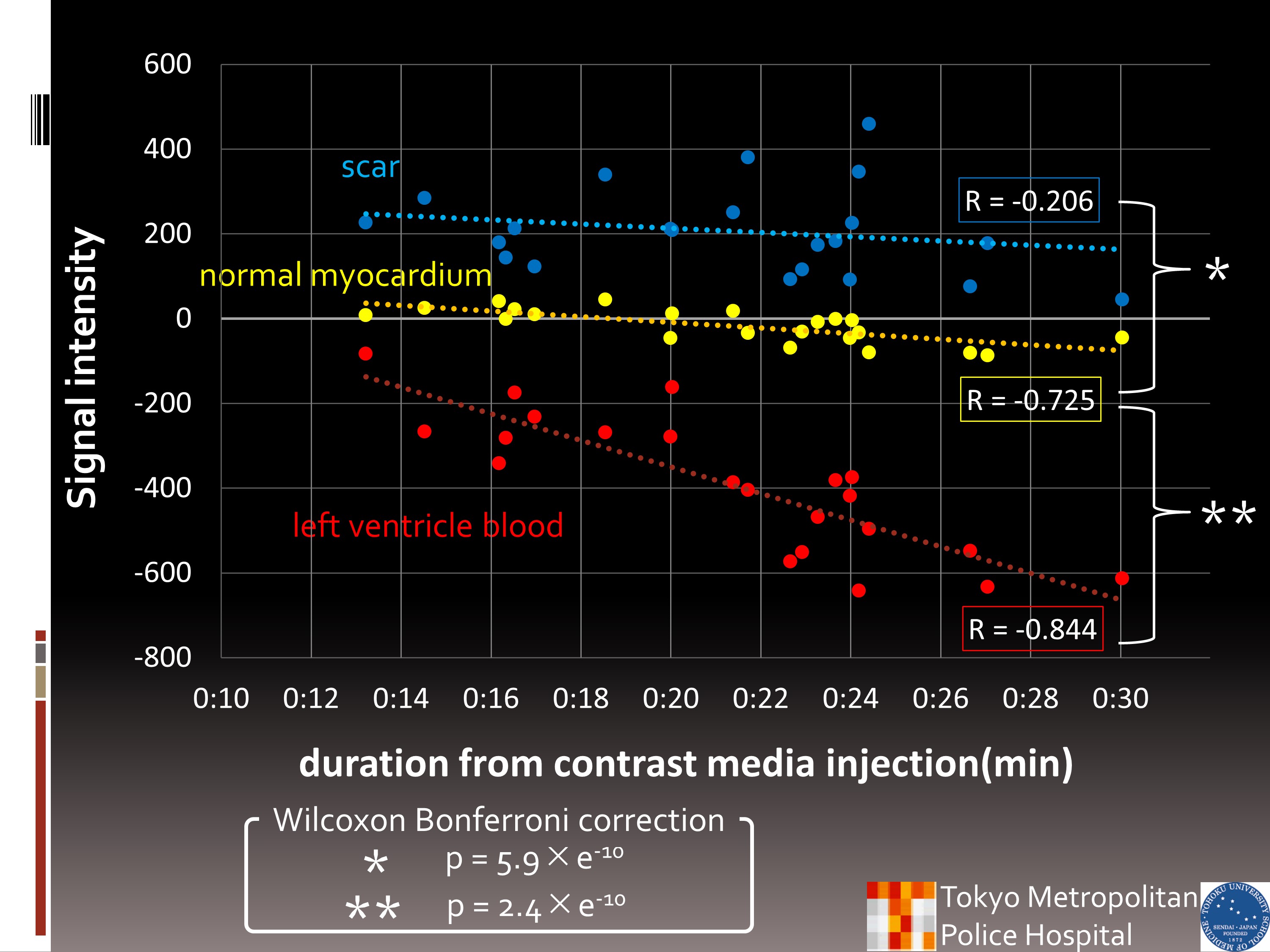
Used MR device was 1.5T Philips Achiva R3.2 system. The scan parameter was set by basic experiments and TI was set shortest (=200ms) for BB Image. Experiment 1: Images of look-locker sequence every 5min after contrast media injection were obtained on 13 volunteers to evaluate signal change of left ventricular blood (LVB) with gadolinium enhancement on PSIR-bSSFP, and to determine the ideal nulling time before BB-LGE imaging. Experiment 2: PSIR-bSSFP Images 10min after the contrast medium injection were obtained on 24 patients with scar. The signal intensities (SIs) of LV blood (LVB), scared myocardium (MYOscar), normal myocardial (MYOnormal) were measured on PSIR-bSSFP images in each patients. And scar volumes of 3D-IR-T1-TFE and those of PSIR-bSSFP were measured.Result
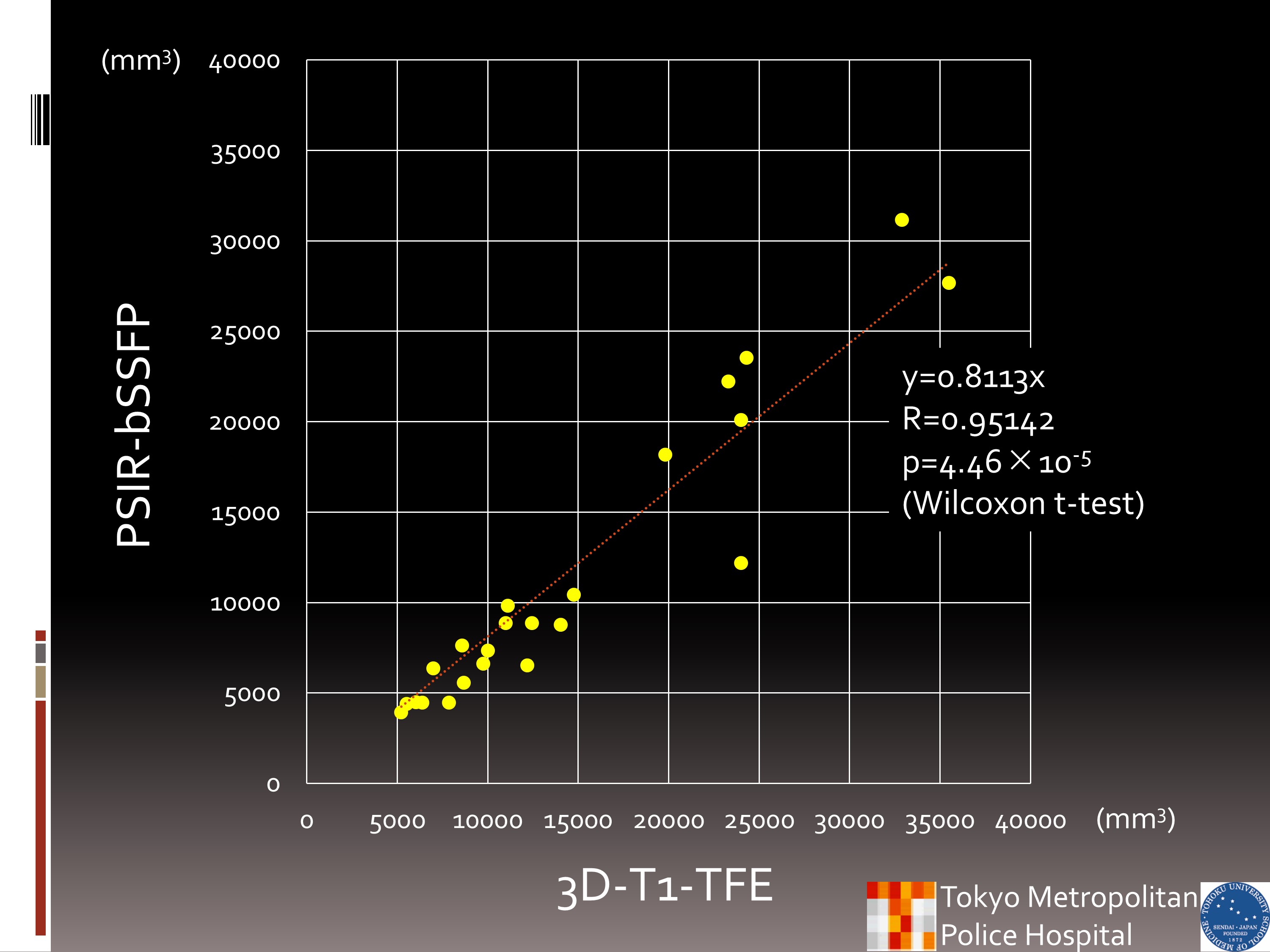
On Ex1, the average nulling time of LVB was longer than 200ms after 10min from injection. The post injection time of “after 10min” on Ex2 was applied by the result Ex1. On Ex2, the signal intensities (SIs) of LVB were negative, those of MYOnormal were zero, and those of scar were positive in all patients’ images. The SIs of LVB and MYOnormal has negative correlation to the duration from contrast injection (LVB:-0.845, MYOnormal:-0.725). However, there was no correlation between the SIs of MYOscar and the duration from gadolinium injection. The SIs among LVB, MYOnormal, and MYOscar were significantly different. The scar volumes were correlated well between images with 3D-IR-T1-TFE and PSIR-bSSFP (r=0.951). The scar volume by PSIR-bSSFP was smaller than that of 3D-IR-T1-TFE.Conclusion
PSIR-bSSFP provide black blood images even after the contrast medium injection, and visualize scared myocardium as high signal intensity area than normal myocardium. The PSIR-bSSFP is one of the sequences that provide good LGE images of myocardial infarction.Acknowledgements
No acknowledgement found.References
(1) Randolph M. Setser, et al. J Magn Reson Imaging 21:650–655 (2005) (2) Jingsi Xie, et al. J Magn Reson Imaging: 32:399–408 (2010) (3) Magalie Viallon, et al. J Magn Reson Imaging: 34:1374–1387 (2011)
Figures