4829
The Value of Magnetic Resonance Black-blood Imaging in Differentiating Acute and Chronic Cerebral Venous Thrombosis1Radiology, Xuanwu Hospital, Capital Medical University, Beijing, China, 2MR Collaboration NEA, Siemens Healthcare, Beijing, China, 3neurosugery, Xuanwu Hospital, Capital Medical University, Beijing, China
Synopsis
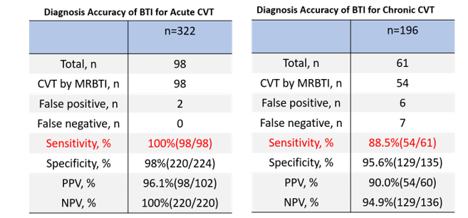
This study aims to demonstrate the value of magnetic resonance black-blood imaging (MRBTI) for differentiating acute and chronic cerebral venous thrombosis (CVT) as well as the diagnosis accuracy of CVT in segment levels. The SNR and CNR of the acute CVT group were significantly higher than that of the chronic group. The sensitivity and specificity of MRBTI were 95.6% (152 /159) and 98.0% (352 /359), respectively. Furthermore, the sensitivity of MRBTI in detecting acute thrombus is up to 100%, compared with 88.5% in the chronic group, which means MRBTI has high sensitivity for early diagnosis.
Purpose
Cerebral venous thrombosis (CVT) is a relatively uncommon disease that often affects young people and pregnant women with untypical symptoms such as headache, vomit and even seizure[1-3]. There are different imaging modalities such as computed tomography (CT), magnetic resonance (MR) used when patients suspected with CVT. However, these traditional techniques only base on blood flow, but not on imaging the thrombus directly. CVT may be misdiagnosed due to the individual differences in venous anatomy. A thrombus in small cortical veins may also be missed due to the low spatial resolution. A newly proposed technique named magnetic resonance black-blood thrombus imaging (MRBTI) has great potential in visualizing the thrombus clearly. The purpose of this study was to evaluate the clinical feasibility of MRBTI in diagnosing different stage of CVT.Methods
MR data was collected on a MAGNETOM Verio 3T MR scanner (Siemens Healthcare, Erlangen, Germany) with a 32-channel head coil. 37 patients (18 male; mean age, 34 years; range, 15-63 years) with CVT diagnosed by routine imaging examinations were enrolled and underwent MRBTI scans. The thrombosed venous sinus were divided into 14 segments (superior sagittal sinus, inferior sagittal sinus, right transverse sinus, right sigmoid sinus, left transverse sinus, left sigmoid sinus, straight sinus, confluence of sinuses, veins of Galen, internal cerebral veins, basal veins of Rosenthal, veins of Labbé, right cortical veins, and left cortical veins). According to the time from onset to MRBTI examination, the patients were divided into an acute group (n=23, ≤14d) and a chronic group (n=14, >15d). The differences of signal-to-noise ratio (SNR) and contrast-to-noise ratio (CNR) between acute and chronic CVT groups were compared using t-test analysis. The magnetic resonance venography (MRV) examination was used as a reference to calculate the accuracy of MRBTI on per-segment level in two groups. The MRBTI images were acquired with a prototype T1-weighted DANTE-SPACE sequence with the following parameters: TR/TE = 650/12 ms, matrix = 272 x 320, FOV = 204 mm x 240 mm (sagittal), slice thickness = 0.75 mm, 208 slices, and total acquisition time = 5:14min.Results
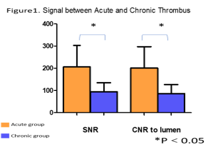
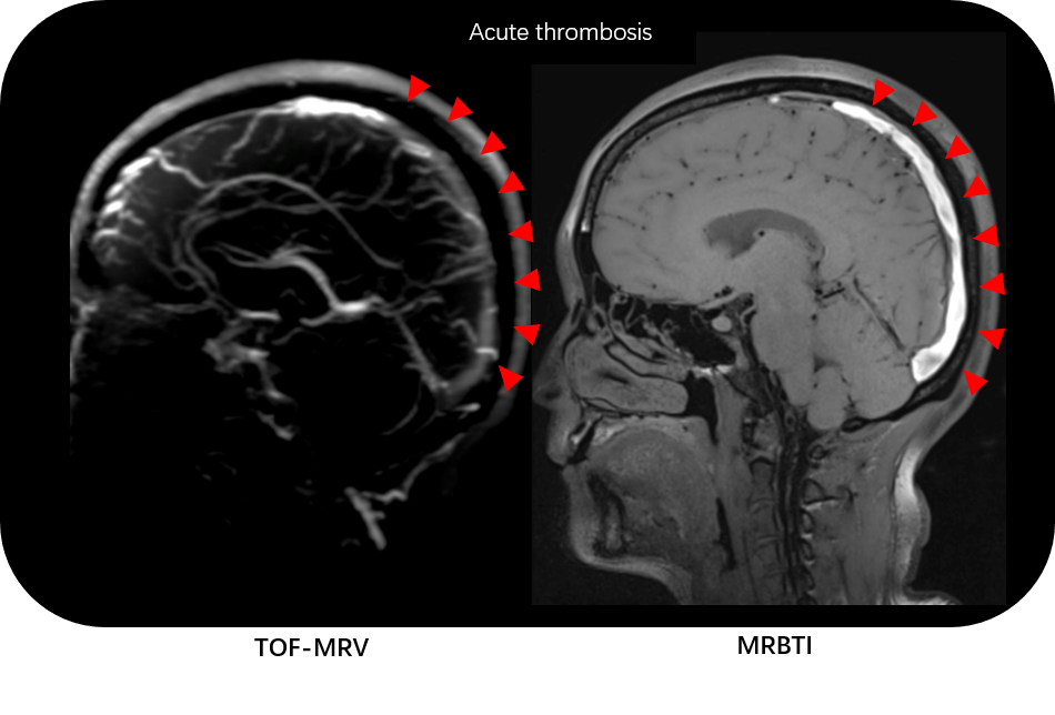
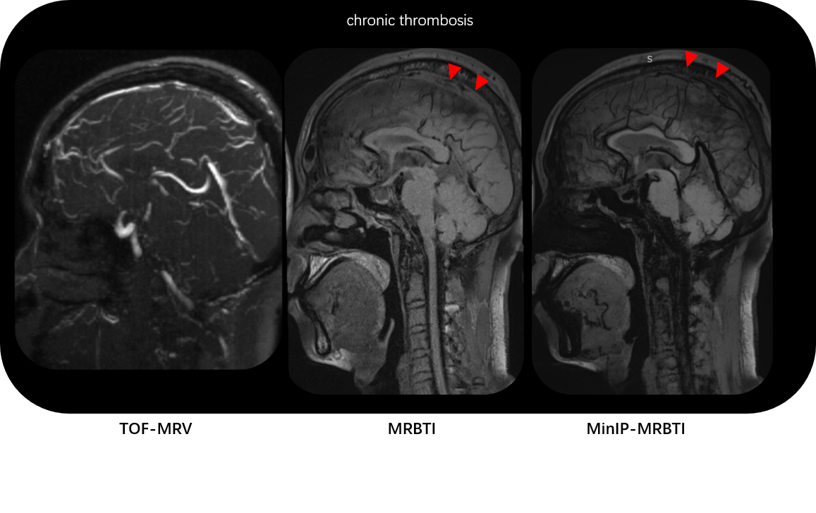
The SNR and CNR of thrombosis in the acute group and chronic group were 206 ± 97 and 94 ± 41, 201 ± 96 and 86 ± 40, respectively. The acute group had significantly higher SNR/CNR than chronic group (t = 4.9/ t = 5.0, P<0.01) (Figure 1). Typical cases of acute and chronic thrombosis are shown in Figure 2 and Figure 3. In 37 patients with CVT, the thrombi in 159 cerebral veins and venous sinus segments were detected with MRV. BTI identified the thrombi accurately in 152 vascular segments, and thrombi in 352 vascular segments could be excluded. The sensitivity and specificity of MRBTI were 95.6% (152 /159) and 98.0% (352 /359) respectively. In the two groups, the diagnosis sensitivity of MRBTI is 100% (98/98) for acute CVT and 88.5% (54/61) for chronic CVT (Table. 2).Discussion
Previous studies have shown that the combination of routine MRI and MRV was helpful in the diagnosis of CVT and allowed it to become the examination of first choice[4]. However, in this study, we found that MRV could not achieve an accurate staging of thrombosis. The MRBTI technique could suppress normal blood signal in the sinus and achieved direct thrombus imaging[5].By this, it could reduce the false positive rate of conventional imaging methods caused by slow blood flow, normal sinus variation, and sinus hypoplasia. Furthermore, we found the diagnostic performance of MRBTI for acute CVT was higher than for chronic CVT. Our results demonstrated that MRBTI could not only improve the accuracy of early diagnosis in CVT, but also show characteristics of variation of hemoglobin signal in acute and chronic phases. This precise staging will contribute to appropriate clinical treatment decision.Conclusion
Achieving direct angiography of cerebral venous thrombosis, the MRBTI technique can accurately differentiate acute or chronic thrombus, which could provide helpful information for treatment decisions.Acknowledgements
No acknowledgement found.References
1.Saposnik, G., F. Barinagarrementeria, R.D. Brown, Jr., et al., Diagnosis and management of cerebral venous thrombosis: a statement for healthcare professionals from the American Heart Association/American Stroke Association. Stroke, 2011. 42(4): 1158-92.
2.Devasagayam, S., B. Wyatt, J. Leyden, et al., Cerebral Venous Sinus Thrombosis Incidence Is Higher Than Previously Thought: A Retrospective Population-Based Study. Stroke, 2016. 47(9): 2180-2.
3.Ferro, J.M., P. Canhao and D. Aguiar de Sousa, Cerebral venous thrombosis. Presse Med, 2016. 45(12 Pt 2): e429-e450.
4.Bonneville, F., Imaging of cerebral venous thrombosis. Diagn Interv Imaging, 2014. 95(12): 1145-50.
5.Xie, Y., Q. Yang, G. Xie, et al., Improved black-blood imaging using DANTE-SPACE for simultaneous carotid and intracranial vessel wall evaluation. Magn Reson Med, 2016. 75(6): 2286-94.
Figures