4828
Quantifying dynamic CBF changes in 4-day follow up after carotid endarterectomy using 3D pCASL1Department of Radiology, Chinese PLA General Hospital, Beijing, China, 2Department of neurosurgery, Chinese PLA General Hospital, Beijing, China, 3GE healthcare China, Beijing, China
Synopsis
Hyperperfusion syndrome was a severe complications of carotid endarterectomy (CEA), the routine measurement for preventing this was to take antihypertensive drugs for at least one week after operative. But there were no definite criteria to assess whether this method was reasonable and appropriate; our objective was to detect dynamic CBF changes in 4-day follow up after CEA using 3D pCASL technique. It is seen that compared to patients before CEA, both the ipsilateral and opposite CBF values were increased in various time points after CEA (P<0.05), whereas, the bilateral CBF values shows no significant differences (P>0.05). This shown that 3D pCASL is sensitive to temporal hemodynamic changes after CEA and may provide a quantitative criterion for assessing antihypertensive drugs taken. Also the observed increases shortly in the first and the second day after operations may suggest for potential drug intervention in case of post-CEA complications.
Purpose
Carotid artery stenosis has high risk for stroke and transient ischemic attack1. Carotid endarterectomy (CEA) is a commonly practiced procedure for treatment of carotid stenosis, especially in patients with symptomatic carotid artery stenosis. It was known that post-CEA hyperperfusion syndrome, one of its severe complications, might result in intracranial hemorrhage and cognitive impairment2. The routine measurement for preventing this was to take antihypertensive drugs for at least one week after operative, but there were no definite criteria to assess whether this method was reasonable and appropriate, thus it was essential to monitor hemodynamics changes after CEA. Our objective was to detect dynamic CBF changes in 4-day follow up after CEA using 3D pseudocontinuous arterial spin labeling (pCASL) technique.Methods and Materials
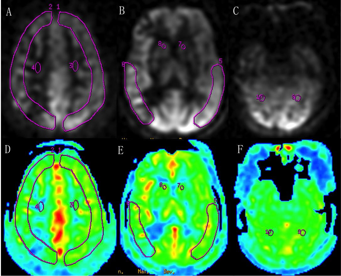
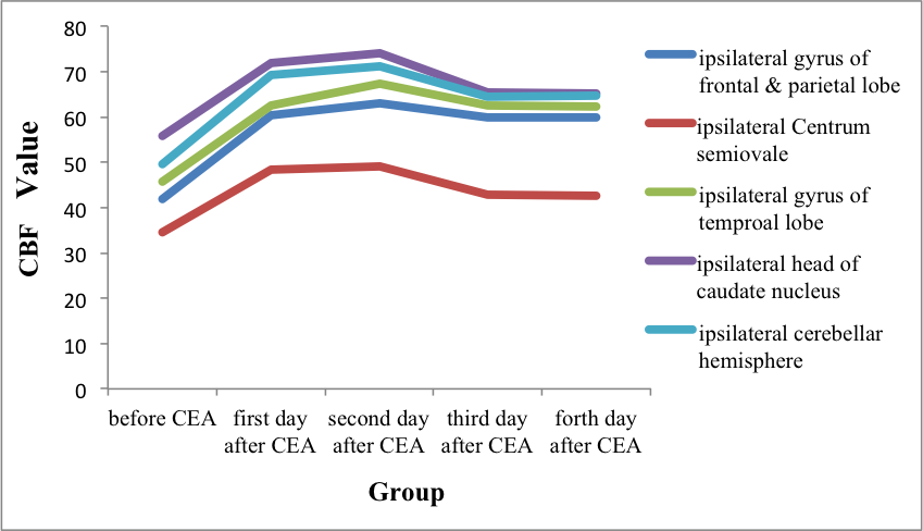
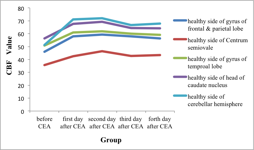
Fourteen patients (male, 11; age, 61.8±9.8 years) were diagnosed with ipsilateral internal carotid artery (ICA) stenosis (≥ 70% narrowing) by neurosurgeon underwent CEA were enrolled in this study, consent forms were obtained. All the participants underwent MR examinations including MRA, DWI and 3D pCASL on a 3.0T whole body scanners at five different time points, including pre-operation, the first day, the second day, the third day and the forth day after operation. Region of interest (ROI) based measurements of CBF data were obtained in several regions of bilateral ICA territory on ASL raw data images, including gyrus of frontal and parietal lobe, centrum semiovale, gyrus of temproal lobe, head of caudate nucleus, and the region of posterior circulation territory, such as cerebellar hemisphere (Figure 1). Repeated measurement ANOVA and paired t-test were performed to compare the significance of the inter-group difference in CBF modifications.Results
The line charts of CBF values at five different time points are shown in Figure 2 and Figure 3. It is seen that the CBF values in ipsilateral of the ICA stenosis increased in the first day (45.5 vs. 62.5, P=0.000), the second day (45.5 vs. 64.37, P=0.000), the third day (45.5 vs. 59.88, P=0.001) and the forth day (45.5 vs. 59.04, P=0.009) after CEA. The opposite CBF values were also increased in the first day (48 vs. 60.07, P=0.008), the second day (48 vs. 61.77, P=0.001), the third day (48 vs. 58.31, P=0.021) and the forth day (48 vs. 58.16, P=0.029) after CEA. No significant difference of CBF was observed at various time points after CEA, also bilateral CBF values showed no statistically significant (P>0.05) inter-group differences (Figure 2, Figure 3).Conclusion
Monitoring the cerebral blood condition after surgical operations for patients with stenosis is of importance for further assessing antihypertensive drugs taken. In this study, it was shown that 3D pCASL is sensitive to temporal hemodynamic changes after CEA and may provide a quantitative criterion for assessing antihypertensive drugs taken. Also the observed increases shortly in the first and the second day after operations may suggest for potential drug intervention in case of post-CEA complications.Acknowledgements
We would like to acknowledge Zhenyu Zhou, Bing Wu, at GE Healthcare China, for their excellent technical assistance.References
1. Eric J. Heyer, Joanna L. Mergeche, E. Sander Connolly Jr. Middle cerebral artery pulsatility index and cognitive improvement after carotid endarterectomy for symptomatic stenosis. J Neurosurg. 2014; 120: 126-131.
2. Xue Z, Peng D, Sun Z, et al. Intraoperative Perfusion Computed Tomography in Carotid Endarterectomy: Initial Experience in 16 Cases. Med Sci Monit, 2016; 22: 3362-3369.
Figures