4796
4D high-resolution Angiography maps obtained by combining low-resolution time-resolved ASL with static high-resolution Time-of-Flight dataThomas Lindner1, Naomi Larsen1, Olav Jansen1, and Michael Helle2
1Department of Radiology and Naeuroradiology, University Hospital Schleswig-Holstein, Kiel, Germany, 2Tomographic Imaging Department, Philips Research Laboratories, Hamburg, Germany
Synopsis
In this study a method to post-process Arterial Spin Labeling (ASL) and Time-of-Flight (TOF) data is presented. The time-resolved low-resolution images of ASL are mapped onto static high-resolution TOF images to create high-resolution time-resolved angiograms.
Introduction
Time-of-flight (TOF) angiography is considered the clinical gold-standard of acquiring non-contrast enhanced angiographic data. The technique generally is acquired as static angiogram [1]. Therefore, information about blood flow is only limited available. Arterial Spin Labeling (ASL) on the other hand can be used for time-resolved (and artery-selective) imaging of blood-flow, yet lacks the spatial resolution that is achievable using TOF angiography [2]. The optimum information about anatomical as well as flow properties would be a method that allows for time-resolved acquisition while keeping the very high resolution of less than 0.5mm in-plane that is achievable using TOF. The aim of this study therefore is to combine these two techniques and make use of the advantages of both TOF and ASL imaging to create high-resolution time-resolved angiograms of the cerebral vasculature.Materials and Methods
Thirteen healthy volunteers underwent MR scanning under the general protocol for sequence development, approved by the local ethical committee. Imaging was performed on a Philips 3T Achieva (Philips, Best, The Netherlands) scanner using a standard 32 channel SENSE Head coil. TOF parameters were: 3D T1-FFE readout with 20° Flip Angle, 0.45*0.45*0.8mm³ voxel size and 135 slices. ASL parameters were: 3D T1-TFE readout with 10° Flip Angle, 0.9*0.9*0.9mm³ voxel size, 120 slices and 6 acquisition time-points with a temporal resolution of 150ms after labeling. To map the time-resolved information from ASL to TOF, at first the ASL data was changed in size using tricubic interpolation in all three axis to match the size of the TOF scan [3]. After image registration, the dynamic ASL data is used as mask for the TOF data, i.e. each time-frame of the ASL is mapped on the TOF image, leading to new TOF images showing different states of blood filling (Figure 2). All image post-processing was performed using Matlab R2013a (The Mathworks, Natick, MA). A flowchart of the post-processing routine is shown in figure 1.Results and Discussion
In all volunteers images could be successfully obtained and used for post-processing. By mapping the temporal information from ASL to the TOF data, it is possible to create an artificial time-resolved TOF image with the additional possibility to remove venous signal contribution that occurs when the field-of-view includes the superior sagittal sinus (Figure 3). The currently obtained images appear well separated regarding their time-points and also provide a good overview of the flow situation within the brain vessels. Furthermore, the technique is not limited to non-selective imaging, but selective ASL angiography can be used accordingly to the here presented method. Using the temporal information of low resolution images (in this case ASL) is a source of uncertainty. Especially the “border zones” of the individual temporal phases of the ASL images need to be further evaluated to accurately show the individual time-points. The accuracy of the here presented combination of these two image types therefore needs to be further evaluated. Combining two individual image sequences poses a time-constraint. Therefore, an acceleration of this technique should be welcomed. This can either be achieved by combining the sequences into a single scan or by reducing scan time especially of the ASL scan by using even lower spatial resolution. How low this resolution of ASL can be set is subject for further investigation.Conclusion
First results of combining time-resolved ASL with TOF imaging appear attractive to obtain time-resolved images of the cerebral vasculature with high spatial resolution.Acknowledgements
No acknowledgement found.References
[1] Saloner D. Radiographics 1995;15:453-465. [2] van Osch MJP et. al. JCBF Metabol 2017; doi: 10.1177/0271678X17713434. [3] Leiken F., Marsden J. Int. J. Numer. Meth. Engng. 2005;63:455–471Figures
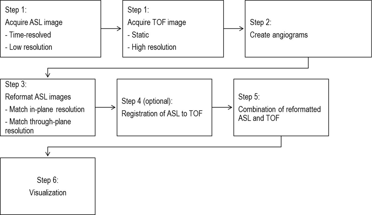
Flowchart of the processing
steps to extract the temporal information from ASL and map it to the TOF scan
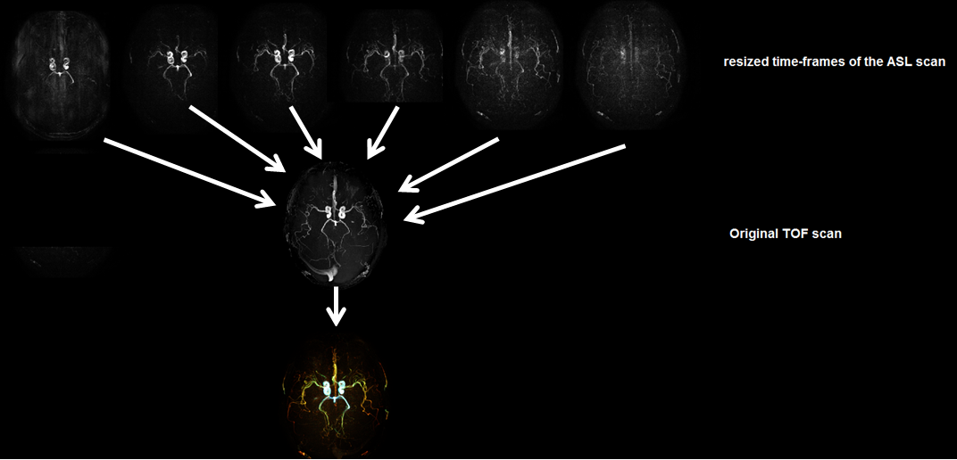
Time-of-arrival
mapping using the information obtained from ASL on the TOF scan.
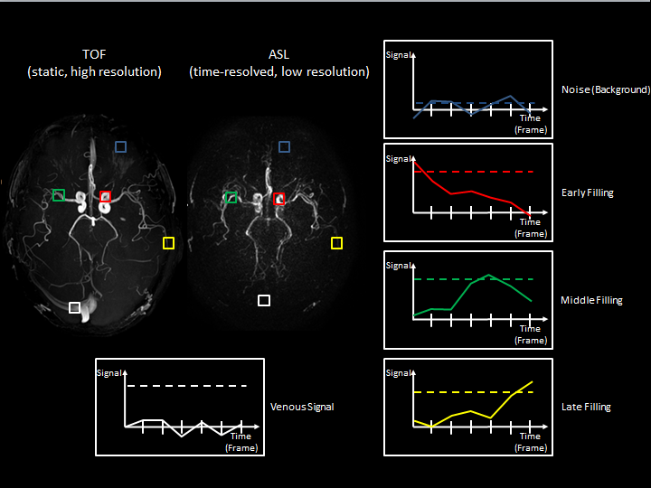
Signal level of ASL
and TOF over time. ASL is shown as solid lines and TOF as dashed lines. ASL
changes its signal levels over time due to flow dynamic behaviour in the
arteries (red, green, yellow). TOF on the other hand is a static imaging
approach, not changing signal levels over time. The white box shows venous
signal which is non-existent in ASL.