4790
Improved motion correction for myocardial T1 map and ECV mapPan ki Kim1, Chul Hwan Park2, Yoo Jin Hong1, and Byoung Wook Choi1
1Department of Radiology and Research Institute of Radiological Science, Severance Hospital, Yonsei University Medical Center, Seoul, Republic of Korea, 2Department of Radiology and Research Institute of Radiological Science, Gangnam Severance Hospital, Yonsei University Medical Center, Seoul, Republic of Korea
Synopsis
Chemical Exchange Saturation Transfer (CEST) has been attracting attention as a molecular imaging method to investigate myocardial muscle energetics according to creatine changes. In this study, we proposed a robust CEST imaging technique from cardiac and respiratory motion using golden angle radial readout to achieve CEST imaging at the heart of the rat. We investigated the feasibility of the proposed method for the creatine phantom and a normal rat.
INTRODUCTION
In cardiac MRI, T1 mapping is applied to a variety of heart diseases and is becoming a valuable diagnostic protocol. Cardiac T1 mapping can be obtained by obtaining a number of heavy T1 weighted images by inversion pulses(IR) during single breath-holding. However, even when a healthy person stops breathing for 10 to 15 seconds, the position of the heart changes slightly due to physiological movements. Therefore, the use of the motion correction method in cardiac T1 mapping can reduce the partial volume effect at the boundary between the myocardium and blood. The motion correction method applied in cardiac T1 mapping may not work well with tissue with short T1 or low contrast between the myocardium and blood.1 In this study, we proposed T1 mapping and ECV mapping method which can provide stable motion correction by providing proper reference image using structure similarity index (SSIM).METHODS
Figure 1 shows the algorithm of proposed motion-correction from T1 weighted images obtained by the MOLLI T1 mapping sequence. First, the initial T1 map is calculated from the first three T1 weighted images affected by the first IR pulse. Using this initial T1 map, it generates synthetic IR weighted images similar to the original image. The original image and the synthetic IR image are evaluated by SSIM, and the highest scored image is set as the reference image and the image based motion correction is performed. The T1 map is calculated from the motion corrected images and the synthetic IR weighted images are reproduced again to perform the motion correction repeatedly. Also, it is possible to automatically generate ECV map using M0 image obtained by calculating T1 map before and after using contrast agent.RESULTS
Figure 2 shows the results when the proposed motion correction method is repeatedly applied. Figure 3 shows the T1 weighted images before and after the motion correction, native T1 and post T1 maps, and the ECV map.DISCUSSION
The Inversion recovery pulses for the T1 mapping generate different contrasts depending on the tissue T1, which makes image-based motion correction difficult. In this case, it is important to provide a reference image with high similarity to the original image for successful motion compensation. As a result, T1 fitting error could be reduced by repetitive calculation as shown in Figure 2. Figure 3 shows that motion correction works well for various IR-weighted images to calculate the T1 map.Acknowledgements
This work was supported by the National Research Foundation of Korea (RF) grant funded by the Korea government (MISP) (No. 2016R1C1B1013837)References
- Xue, Hui, et al. "Motion correction for myocardial T1 mapping using image registration with synthetic image estimation." Magnetic resonance in medicine 67.6 (2012): 1644-1655.
Figures
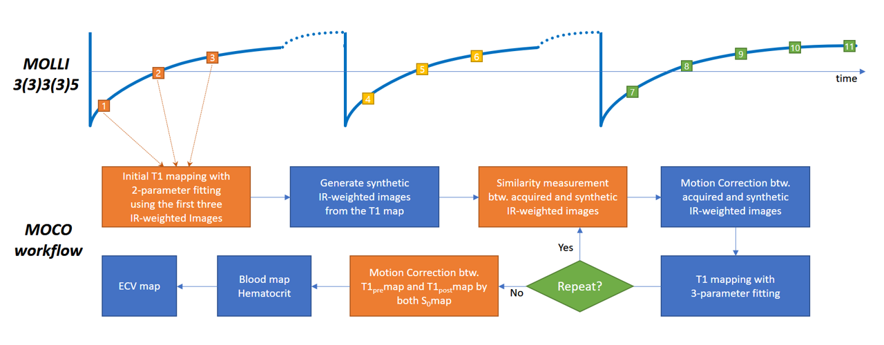
Motion
correction workflow for myocardial T1 mapping
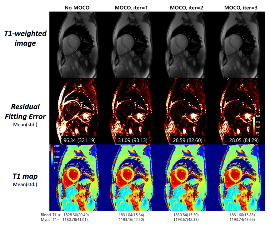
The
result of repeatedly applying the proposed motion compensation
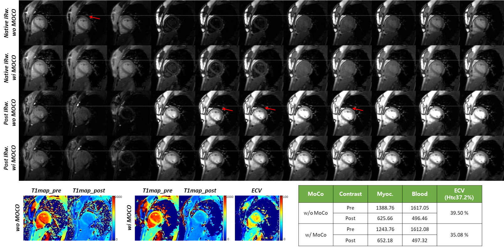
T1
map and IR weighted images before and after applying proposed motion
compensation