4787
Deep Learning Image Classification for Automatic Frequency Adjustment in High Field Cardiac MR Imaging1Research and Education, St Francis Hospital, Roslyn, NY, United States
Synopsis
Deep learning using a 3D CNN is a viable solution for the determination of the center frequency at high fields for cardiac imaging. It can quickly provide similar results to an expert observer. In the future, the strategy may be applicable to improved frequency resolution and to frequency adjustments outside of the frequency scout range. This strategy should make 3T cardiac MR imaging accessible to a wider audience.
Introduction
Cardiac bSSFP CINE imaging at magnetic fields above 1.5T remains very sensitive to the setting of the adjusted frequency. Suboptimal setting of this frequency causes severe banding, flow artifacts and artifactual signal shifts. These artifacts can often render images unsuitable for clinical decisions. The current solution is to obtain several images over a range of frequencies (a so-called frequency scout) and select an optimal frequency by visually evaluating the images. Although this technique can be effective, there are several drawbacks. Firstly, it requires additional technologist training beyond the typical cardiac MR training. Secondly, the decision is time consuming because the optimal frequency varies significantly across imaging planes (HLA, VLA, LVOT and short-axis). Additionally, the decision can be quite subjective. The observer must pick a frequency optimal across the entire heart while excluding artifacts from implanted metal and flow. The purpose of this study was to develop and evaluate a deep learning approach for automatic cardiac MR frequency tuning at high fields.Methods
This study was performed at 3T with a clinical Siemens Skyra imager. Frequency scouts from 100 patients referred for clinical evaluation were included in this IRB approved study; including 4 anatomical slice planes (mid short-axis, two-, three-, and four-chamber views). Thirteen images were obtained with frequencies ranging from -150 to 150 Hz with a 25 Hz increment. The 2D bSSFP frequency scout sequence had parameters matching CINE imaging: TR/TE= 2.5/1.25 ms, bandwidth 1095 Hz/pixel and slice thickness=8 mm. A 3D linear convolutional neural network (CNN) was used to determine the optimal frequency. The problem is three-dimensional in nature (two in-plane dimensions and one frequency dimension) Keras v2.0 and TensorFlow v1.3 (two open-source software libraries) were used for deep learning implementation. 200 image sets were used for 3D CNN training. The remaining 200 images sets were used for optimal selection of frequency. The performance of the new method was evaluated with Pearson correlation when comparing manual and automated frequency determination.Results
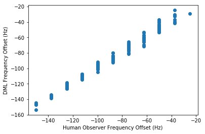
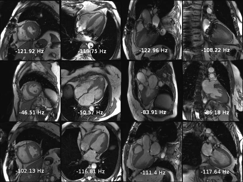
Training of the deep learning algorithm was performed in 50 epochs with excellent validation accuracy (Figure 1, R2=0.99). Representative examples of the prospective application of the method are displayed in Figure 2. The frequency offset variance across subjects and imaging planes is well demonstrated. Additionally, the performance in subjects with artificial heart valves and sternal wires was excellent (Figure 2, top and bottom rows). Subsequent prospective CINE imaging in subjects yielded excellent results.without banding artifacts.Conclusion
Deep learning using a 3D CNN is a viable solution for the determination of the center frequency at high fields for cardiac imaging. It can quickly provide similar results to an expert observer. In the future, the strategy may be applicable to improved frequency resolution and to frequency adjustments outside of the frequency scout range. This strategy should make 3T cardiac MR imaging accessible to a wider audience.Acknowledgements
No acknowledgement found.References
1. Deshpande VS, Shea SM, Li D. Artifact reduction in true-FISP imaging of the coronary arteries by adjusting imaging frequency. Magn Reson Med 2003;49(5):803-809.
2. Wansapura J, Fleck R, Crotty E, Gottliebson W. Frequency scouting for cardiac imaging with SSFP at 3 Tesla. Pediatr Radiol 2006;36(10):1082-1085.
Figures