4753
Surveillance of abdominal aortic aneurysm using accelerated 3D non-contrast black-blood MRI with compressed sensing (CS-DANTE-SPACE)1Radiology, University of California, San Francisco, San Francisco, CA, United States, 2Radiology, Xuanwu Hospital, Beijing, China, 3Radiology, Anzhen Hospital, Beijing, China, 4Siemens Healthcare, San Francisco, CA, United States, 5Siemens Healthcare, Erlangen, Germany
Synopsis
Although 3D non-contrast high-resolution black-blood MRI (DANTE-SPACE) is a promising tool for the surveillance of abdominal aortic aneurysm (AAA), it requires lengthy scans (~7 minutes). We implemented a compressed sensing method (CS-DANTE-SPACE) to reduce the scan time by 41% (to ~4 minutes), and tested its feasibility in 20 AAA patients undergoing routine follow-up. We found CS-DANTE-SPACE achieved accurate diameter/area measurements and intraluminal thrombus (ILT) identification, and provided better contrast and vessel sharpness. CS-DANTE-SPACE is a promising tool for AAA surveillance in the clinical setting.
Introduction
Abdominal aortic aneurysm (AAA) is a common condition and is associated with high mortality. Currently, clinical management of AAA is based on the maximal diameter, and AAAs with diameter >5.5cm are candidates for intervention. The majority of patients with smaller AAAs (<5.5cm) are followed with surveillance programs. We previously developed a 3D non-contrast black-blood MRI technique (DANTE-SPACE 1) for use in AAA surveillance, that showed good accuracy and reproducibility compared to gold-standard CTA2. However, this technique requires a scan of ~7 minutes. Scans of this length are poorly tolerated by patients and are more vulnerable to patient motion. This study aims to evaluate whether an accelerated DANTE-SPACE technique using compressed sensing (CS-DANTE-SPACE) can be used for surveillance of patients with AAA disease with the benefit of scan time reduction.Methods
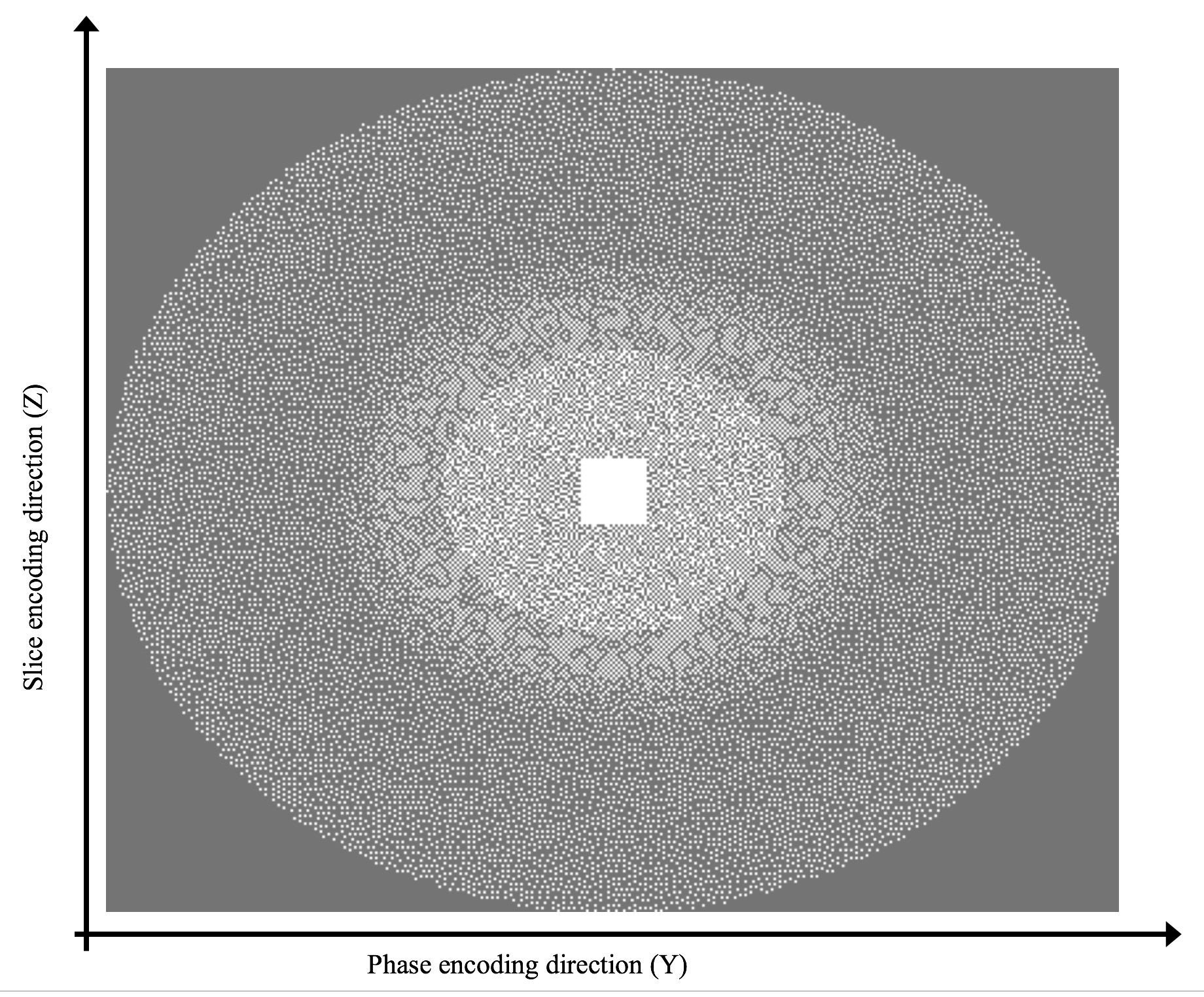
Study population: 20 AAA patients (all male, age 69±8 years) under clinical surveillance were recruited for this study. This is a part of a clinical trial that follows up with AAA patients every 6 months using high-resolution MRI. 2 patients were scanned again 6 months later. In total, 22 image datasets were acquired. Sequences: MR imaging was performed on a clinical 3T system (MAGNETOM Skyra, Siemens Healthcare, Erlangen, Germany). All patients underwent T1-weighted DANTE-SPACE (prototype) and CS-DANTE-SPACE (prototype) scans. Scanning parameters: TR/TE 800/20ms; echo train length 60; 1.3mm isotropic resolution. For DANTE-SPACE, parameters were; GRAPPA 2, 6/8 partial Fourier, avg=3.4 (multiple averages were used to average abdominal motion and improve SNR1), and scan time= 7:10. For CS-DANTE-SPACE, parameters were; a variable-density Poisson disk pattern (Figure 1) with 20 % under-sampling, avg=4, and scan time= 4:12min (59% of DANTE-SPACE) A regularization factor of 0.002 and 20 iterations were used for image recon 3. Image Analysis: Two radiologists, blinded to the sequences used, independently read images and measured the maximal diameter of AAA, the intra-luminal thrombus (ILT)/wall and lumen area, ILT-to-muscle signal intensity ratio, and the ILT/wall-to-lumen contrast ratio. One reviewer measured the inner lumen boundary and outer wall boundaries’ sharpness in two orthogonal directions across the aneurysm (left to right and anterior to posterior) using a previously defined method 4.Results
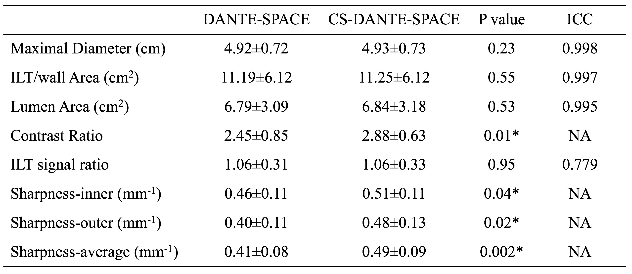
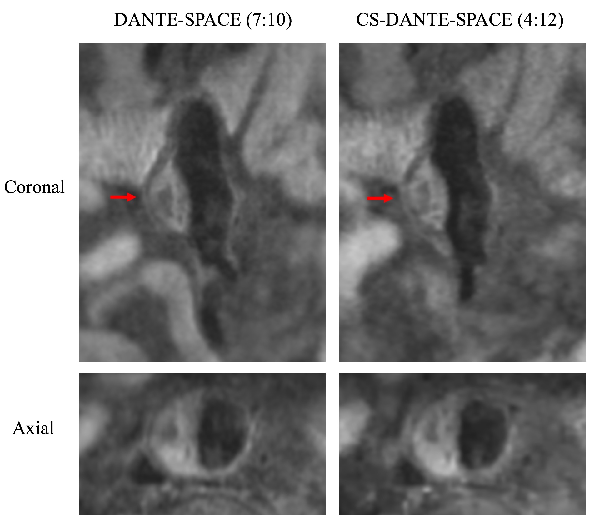
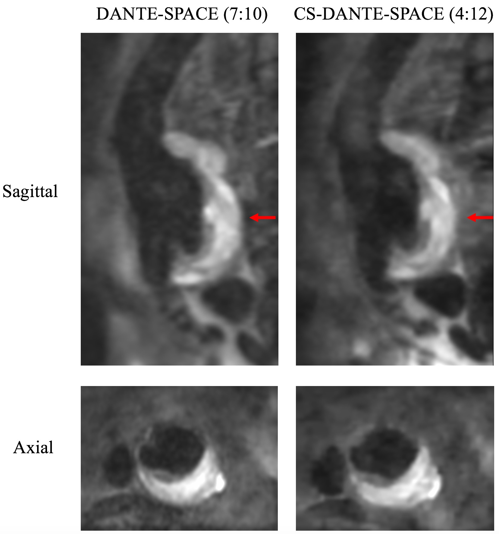
Quantitative comparison of the two sequences is shown in Table 1. Sample images of DANTE-SPACE and CS-DANTE-SPACE are shown in Figure 2 and 3. There was excellent agreement between the two sequences for diameter and area measurements (ICCs>0.99) and good agreement for ILT ratio measurements (ICC = 0.779). There was no significant bias between the sequences (p>0.05). CS-DANTE-SPACE had a higher ILT/wall-to-lumen contrast ratio (p=0.01) and higher lumen/wall boundary sharpness compared with DANTE-SPACE (p=0.002). Results from the two radiologists’ reading were in excellent agreement for diameter/area measurements (ICC = 1.000 and 0.998) and ILT signal ratio measurements (ICC = 0.941).Discussion
This study demonstrated that the compressed sensing method can be used in a clinical setting for the routine follow-up with patients with AAA condition. To our best knowledge, this is the first study showing that compressed sensing black-blood vessel wall imaging can be used in a clinical surveillance program. Previous studies were limited in that they only included healthy volunteers or a very small number of patients (n<10)5 6. Even with a reduction of 41% in scan time, higher ILT/wall-to-lumen contrast ratio, and better sharpness, CS-DANTE-SPACE achieved diameter/area measurements of the AAAs that were comparable to that of conventional DANTE-SPACE. The improved sharpness and contrast ratio found for CS-DANTE-SPACE is likely related to decreased breathing artifacts and bulk motion that result from the reduction in scan time. This is apparent in Figure 2. Considering that AAA patients often have an age >65 years, a reduction in scan time can improve patient comfort and increase the success rate of scanning. In AAA surveillance programs, repeated scans are regularly performed, and a reduction of failed studies also reduces monitoring costs. CS-DANTE-SPACE also reliably quantifies the ILT signal which is significant as the ILT composition is an emerging biomarker for AAA progression7.Conclusion
Compressed sensing black-blood MRI (CS-DANTE-SPACE) can reduce scan time while maintaining image quality in AAA imaging. It is a promising tool for the surveillance of patients with AAA disease in the clinical setting.Acknowledgements
No acknowledgement found.References
1. Zhu C, Haraldsson H, Faraji F, Owens C, Gasper W, Ahn S, et al. Isotropic 3d black blood mri of abdominal aortic aneurysm wall and intraluminal thrombus. Magnetic resonance imaging. 2015
2. Zhu C, Tian B, Leach JR, Liu Q, Lu J, Chen L, et al. Non-contrast 3d black blood mri for abdominal aortic aneurysm surveillance: Comparison with ct angiography. Eur Radiol. 2017;27:1787-1794
3. Stalder AF, Schmidt M, Quick HH, Schlamann M, Maderwald S, Schmitt P, et al. Highly undersampled contrast-enhanced mra with iterative reconstruction: Integration in a clinical setting. Magn Reson Med. 2015;74:1652-1660
4. Zhu C, Haraldsson H, Tian B, Meisel K, Ko N, Lawton M, et al. High resolution imaging of the intracranial vessel wall at 3 and 7 t using 3d fast spin echo mri. MAGMA. 2016;29:559-570
5. Yuan J, Usman A, Reid SA, King KF, Patterson AJ, Gillard JH, et al. Three-dimensional black-blood multi-contrast carotid imaging using compressed sensing: A repeatability study. Magma. 2017
6. Makhijani MK, Balu N, Yamada K, Yuan C, Nayak KS. Accelerated 3d merge carotid imaging using compressed sensing with a hidden markov tree model. Journal of magnetic resonance imaging : JMRI. 2012;36:1194-1202
7. Nguyen VL, Leiner T, Hellenthal FA, Backes WH, Wishaupt MC, van der Geest RJ, et al. Abdominal aortic aneurysms with high thrombus signal intensity on magnetic resonance imaging are associated with high growth rate. Eur J Vasc Endovasc Surg. 2014;48:676-684
Figures