4717
Direct visualization of physiological intestinal flow using unenhanced MR imaging with spin labeling1Asahi General Hospital, Asahi, Japan, 2University of California San Diego, San Diego, CA, United States, 3Toshiba Medical Systems Corp, Tokyo, Japan, 4Hasuda Hospital, Hasuda, Japan
Synopsis
Assessment of motility and propagation of the small bowel has been reported using conventional contrast x-ray techniques with unavoidable ionizing radiation and recent MR imaging. However, MR enteroclysis and enterography require a discomforted nasoenteric intubation and an oral administration of large volumes of enteric contrast materials, respectively. Unenhanced MR imaging with a spin labeling technique provides direct visualization of physiological intestinal intraluminal flow related to bowel peristalsis.
Purpose
Direct monitoring of intestinal content movement related to bowel peristalsis is extremely challenging. Conventional contrast x-ray techniques allowed a measurement of small bowel flow using ionizing radiation. Motility of the small bowel wall with its contractions or distentions has been examined with MR imaging. However, MR enteroclysis requires an administration of contrast material through a nasoenteric tube, whereas MR enterography requires large volumes of enteric contrast materials orally (1, 2). There are few studies investigating the small bowel flow in humans (3). The aim of our study is to directly visualize physiological intestinal intraluminal flow related to the peristalsis without enteric contrast administration by using an unenhanced MR imaging with a spin labeling technique.
MATERIALS AND METHODS
A flow-out technique of time-spatial labeling inversion pulse (time-SLIP) technique was applied on move-out regions by using both a nonselective inversion recovery pulse and a spatially selective inversion labeling pulse. We set the region of interest on the duodenum because of less migration due to retroperitoneal fixation. A protocol optimization was performed on 20 healthy volunteers. All the studies were performed on a 3T MRI system equipped with Atlas SPEEDER body coil. Principal parameters are: respiratory-gated FASE sequence with a single-shot two-dimensional time-SLIP technique during a total of 165-175 seconds (total of 50 images), TR/TE = 3300-3500/80 ms, TI = 1400 ms, matrix = 224×256, FOV = 35 cm×30 cm, parallel imaging factor = 2, slice thickness = 35mm, labeled pulse width = 60mm. All images were evaluated by two radiologists to assess depiction of duodenal content movement. MR peristalsis imaging was evaluated for the presence, frequency, and magnitude of bowel content movement in move-out areas.RESULTS
MR peristalsis imaging with the time-SLIP flow-out technique provided an excellent dynamic imaging in physiological conditions of the duodenum. The tagged intestinal content if not contains luminal gas was satisfactorily visualized after applying the pulse labeling. The intestinal fluid inflow was observed at 8-24 times (mean 12.5 times) in a series of 50 images. The distance that the intestinal fluid moved in the duodenum within the tagged area was 0.5-8.5 cm (mean 3.3 cm) during 1400ms (black blood traveling time). The results of velocity and frequency of the intestinal flow was compatible within the previously reported range on radio-fluoroscopy in normal variants (4).CONCLUSION
Noninvasive visualization of the physiologic intestinal flow is possible by using the unenhanced MR imaging with the time-SLIP flow-out technique. We can also apply this technique to beverage or drug-induced intestinal motility changes.Acknowledgements
No acknowledgement found.References
- Masselli G. MR imaging of the small bowel. Radiology 2012;264:333-348.
- Froehlich JM. Small bowel motility assessment with magnetic resonance imaging. JMRI 2005;21:370–375.
- Gutzeit A. Feasibility of small bowel flow rate measurement with MRI. JMRI 2010;32:345-351.
- Flickenstein P. Electrical spike activity in the human small intestine. A multiple electrode study of fasting diurnal variations. Am J Dig Dis 1978;23:776–780.
Figures
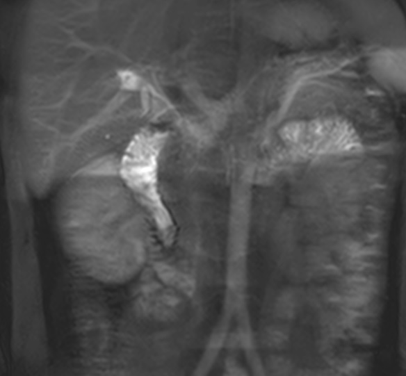
Fig. 1) Unenhanced time-SLIP MR imaging of intestinal flow with a spatially selective IR pulse applied on the upper duodenum.
One peristalsis allowed duodenal fluid to flow into the descending part.
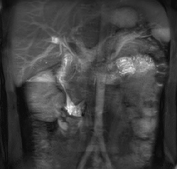
Fig. 2) Unenhanced time-SLIP MR imaging of intestinal flow with a spatially selective IR pulse applied on the upper duodenum.
Another peristalsis allowed duodenal fluid to flow into the horizontal part.