4709
Evaluation of Extracellular Volume (ECV) Fraction of the Normal Pancreas and Correlation with Biophysical Parameters1Radiology and Imaging Sciences, Indiana University School of Medicine, Indianapolis, IN, United States, 2Indiana University School of Medicine, Indianapolis, IN, United States
Synopsis
ECV fraction can be a very useful imaging tool for non-invasive evaluation of solid organ pathologies and is probably underutilized in abdominal imaging. There are potentially several clinical applications of ECV in the abdomen such as evaluation of chronic pancreatitis or chronic liver disease. In this study, we computed the ECV fraction of the pancreas in 60 healthy cohorts and determined that ECV fraction of the normal pancreas is 0.26 (± 0.08). ECV fraction is not influenced by pancreatic steatosis or patient’s gender, however gradually increases with age.
INTRODUCTION
Extracellular volume (ECV) fraction, which requires obtaining T1 relaxation times in pre- and post-contrast phases in both the tissue and inflowing arterial blood has been successfully used as a biomarker for tissue fibrosis, but mostly for myocardial imaging (1). Pancreas is one of the organs that quantitative imaging such as T1 mapping and ECV fraction can be helpful for certain pancreatic pathologies most notably fibrosis seen in chronic pancreatitis (2). There has been no prior study that reported the ECV fraction of the pancreas. Purpose of this study was to determine the normal ECV fraction of the pancreas in patients with no pancreas disease and correlate with age, gender, fat signal fraction (FF), and diameter of the gland.MATERIALS AND METHODS
This HIPAA compliant study was performed on 60 patients who were being screened for pancreatic cancer due to genetic predisposition however, otherwise were healthy individuals. Patient’s history, laboratory, endoscopic ultrasound and Magnetic Resonance Cholangiopancreatography (MRCP) results showed no evidence of acute findings and Cambridge classification of the pancreatic ducts was 0 (normal) (3). All the patients were imaged on a 3T MR scanner (MAGNETOM Verio, Siemens Healthcare GmbH, Erlangen, Germany) using the same imaging protocol which included T1 mapping using dual flip angle VIBE sequence for obtaining the ECV fraction. T1 maps were acquired in pre-contrast and 6-minute late enhancement phase following injection of gadobenate dimeglumine using the manufactured recommended dose of 0.1 mmol/kg. T1 maps were reconstructed online at the MR scanner via vendor supplied software (MapIt, Siemens Healthcare GmbH, Erlangen, Germany). Regions of interest were drawn on the T1 maps over the aortic lumen and pancreas. These measurements were used to generate ECV maps using the formula at each pixel: ECV= [1–hematocrit] x ΔR1pancreas / ΔR1blood, where ΔR1 is the difference of pre- and post-contrast T1 relaxivity. FF of the pancreas was calculated by measuring signal intensity (SI) on the axial breath-hold 2-point DIXON images using the formula FF = SI Fat / [SI Fat + SI Water] (4). Pearson correlation coefficient and two-tailed probability t-test were used to assess the relationship between the ECV and biophysical parameters.
RESULTS
Patients in this study were between 19-78 years of age (mean: 49.8). The mean ECV fraction was 0.26 (SD ± 0.08), FSF was 0.07 (SD ±0.03) and the average diameter was 24 mm (SD ±3.9 mm). Mean diameter was 23.3 mm (SD ± 2.8) in females vs 25.3 mm (SD ±3.4) in males (p=0.04).
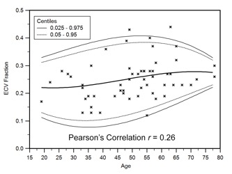
Mean ECV in females was 0.26 (SD: ±0.08) which was not significantly different than the males 0.25 (SD: ±0.07) (p=0.89). ECV showed a weak positive correlation with age (r= 0.26, p=0.04) (Figure 1), but no correlation with FSF (r=0.001, p=0.99). Age showed a moderate negative correlation with diameter (r= - 0.52, p<0.0001) and a weak positive correlation with FSF (r=0.24, p=0.06).
CONCLUSION
The mean ECV fraction of the pancreas is 0.26 (SD ± 0.08). There is a weak positive correlation between ECV fraction with patient’s age. ECV is not influenced by gender or pancreatic steatosis.Acknowledgements
No acknowledgement found.References
1. Schelbert EB, Messroghli DR. State of the Art: Clinical Applications of Cardiac T1 Mapping. Radiology 2016; 278:658-676
2. Tirkes T, Lin C, Fogel EL, Sherman SS, Wang Q, Sandrasegaran K. T1 mapping for diagnosis of mild chronic pancreatitis. Journal of magnetic resonance imaging : JMRI 2017; 45:1171-1176
3. Sarner M, Cotton PB. Classification of pancreatitis. Gut 1984; 25:756-759
4. Cassidy FH, Yokoo T, Aganovic L, et al. Fatty liver disease: MR imaging techniques for the detection and quantification of liver steatosis. Radiographics : a review publication of the Radiological Society of North America, Inc 2009; 29:231-260
Figures