4698
Evaluation of Uterine and Placenta Motion throughout Early Gestation1Radiological Sciences, University of California, Los Angeles, Los Angeles, CA, United States, 2Biomedical Physics, University of California, Los Angeles, Los Angeles, CA, United States, 3Department of Epidemiology, University of California, Los Angeles, Los Angeles, CA, United States, 4Department of Pediatrics, University of California, Los Angeles, Los Angeles, CA, United States, 5Department of Obstetrics and Gynecology, University of California, Los Angeles, Los Angeles, CA, United States
Synopsis
When imaging the uterus and placenta, in pregnant patients, there are potential uterine contractions that compress the superior region of the uterus and can cause significant motion in the uterus and placenta. It is not well studied how much uterine contraction and other motion need to be accounted for during MRI scans in early gestation. In this study we observed and characterized uterine contractions and other bulk motion in pregnant women (gestational age 14-22weeks) using an image based template-matching program. This study can further help develop proper scanning protocols for MRI studies with pregnant patients to avoid potential motion artifacts.
Introduction
Motion during MRI scanning continues to be a major limiting factor that causes imaging artifacts that degrade image quality and quantification. When imaging the uterus and placenta there is maternal respiratory motion, fetal motion, and uterine contractions that need to be considered1,2. In particular, uterine contractions compress the superior region of the uterus and can cause significant motion in the uterus and placenta. However, it is not well studied how much uterine contraction motion and other motion need to be accounted for during MRI scans in early gestation. In this study we evaluate uterine contraction motion and other uterine and placental bulk motion in early gestation (2nd trimester) by using image based motion tracking from dynamically reconstructed golden radial gradient echo images.Methods
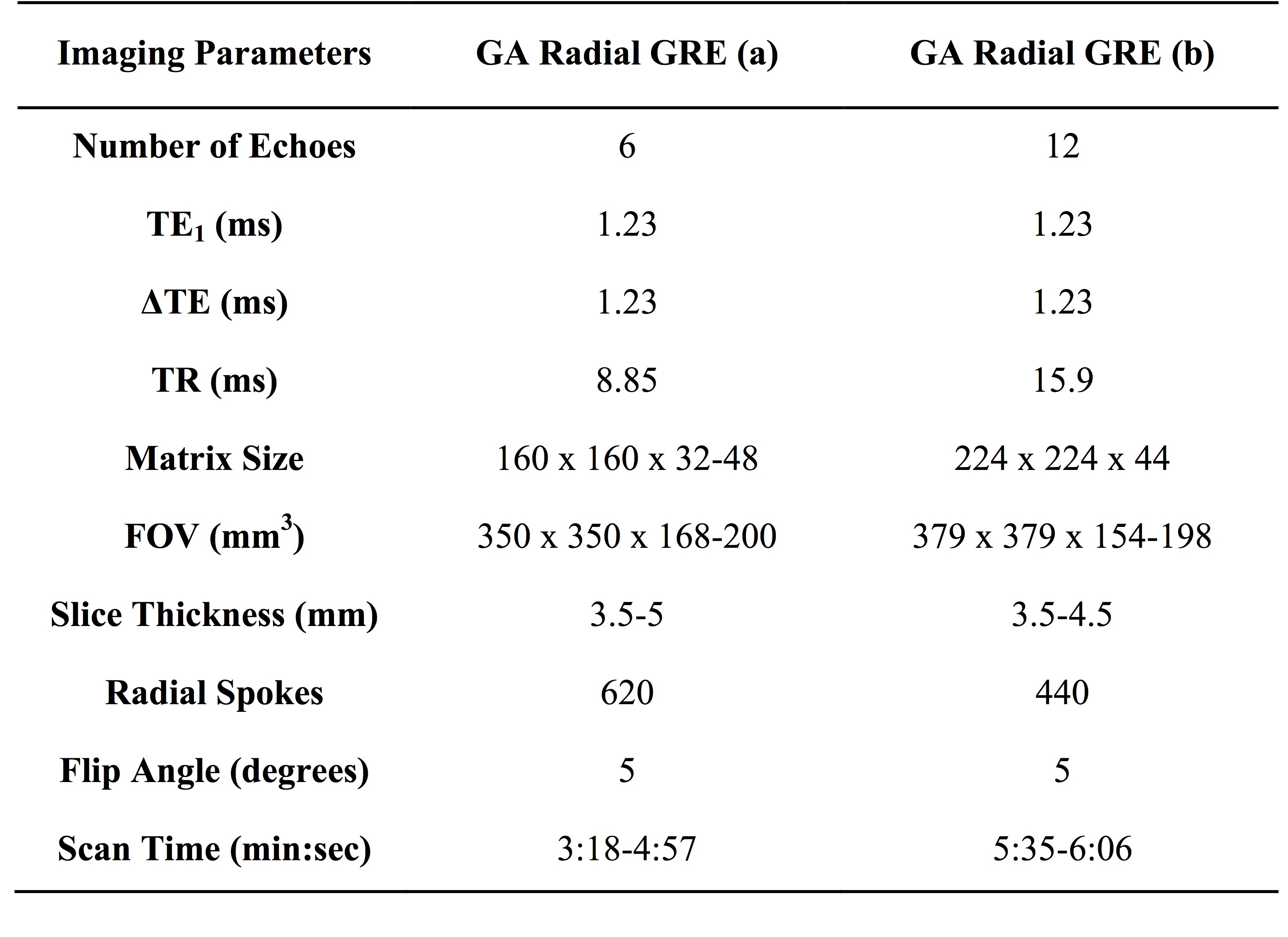
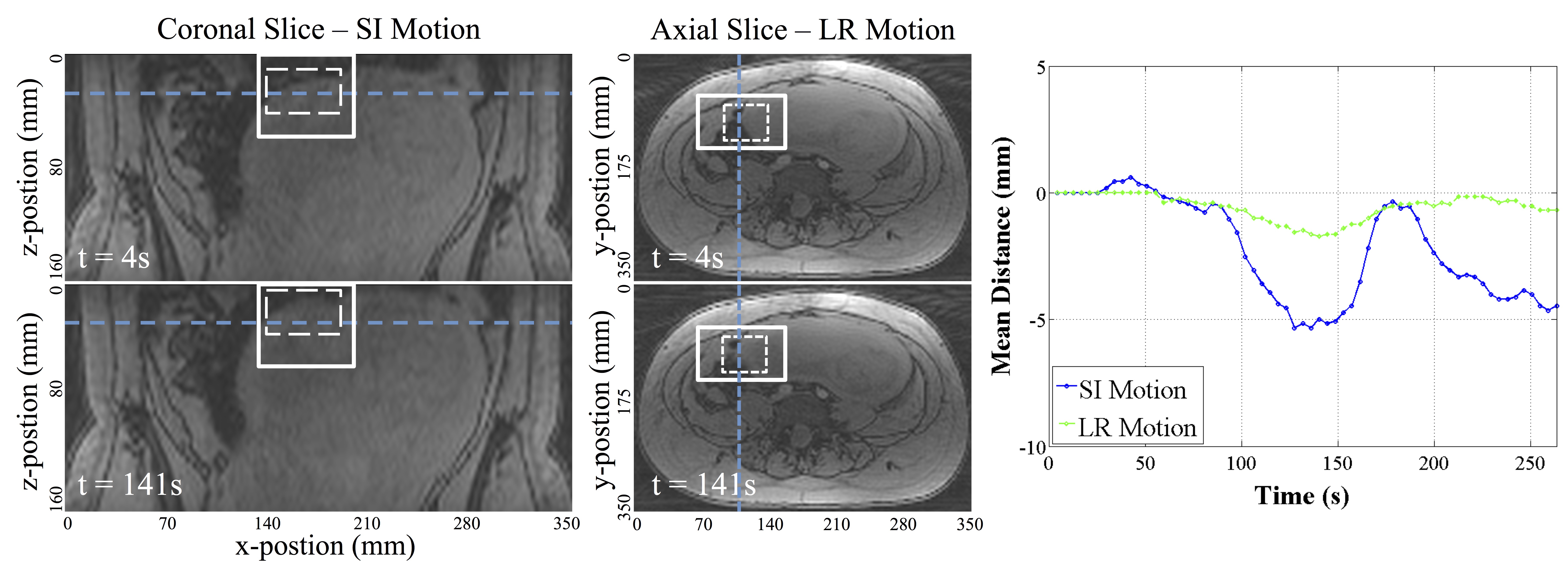
Fifty-one pregnant women were enrolled in a placental function and fetal outcome study. Of these subjects twenty-six were randomly selected to assess uterine contraction and other bulk motion of the uterus and placenta. Each subject was scanned twice at two different time points during their second trimester (first scan 14-16 weeks, second scan 19-22 weeks) for a total of 52 scans. A 3D multi-echo golden angle (GA) radial GRE sequence was used for data acquisition, which was then reconstructed offline using k-space weighted image contrast (KWIC)3 to create high spatial resolution dynamic image series without much streaking. There were two main protocols used for imaging and are listed in figure 1. The first echo was used for reconstruction as that had the highest signal. Motion in the superior/inferior (SI) and left/right (LR) directions was measured using an image based template-matching algorithm4 (see Fig 2). The maximum amplitude of the contraction or bulk motion during the scan was used. The duration of each of the contractions was also measured. Lastly, the area under the curve (AUC) for all contractions cumulatively, and for each contraction, was measured. To assess the dominant magnitude and direction of contraction and other bulk motion in the LR and SI directions, the singular value decomposition (SVD) was used to determine singular values.Results
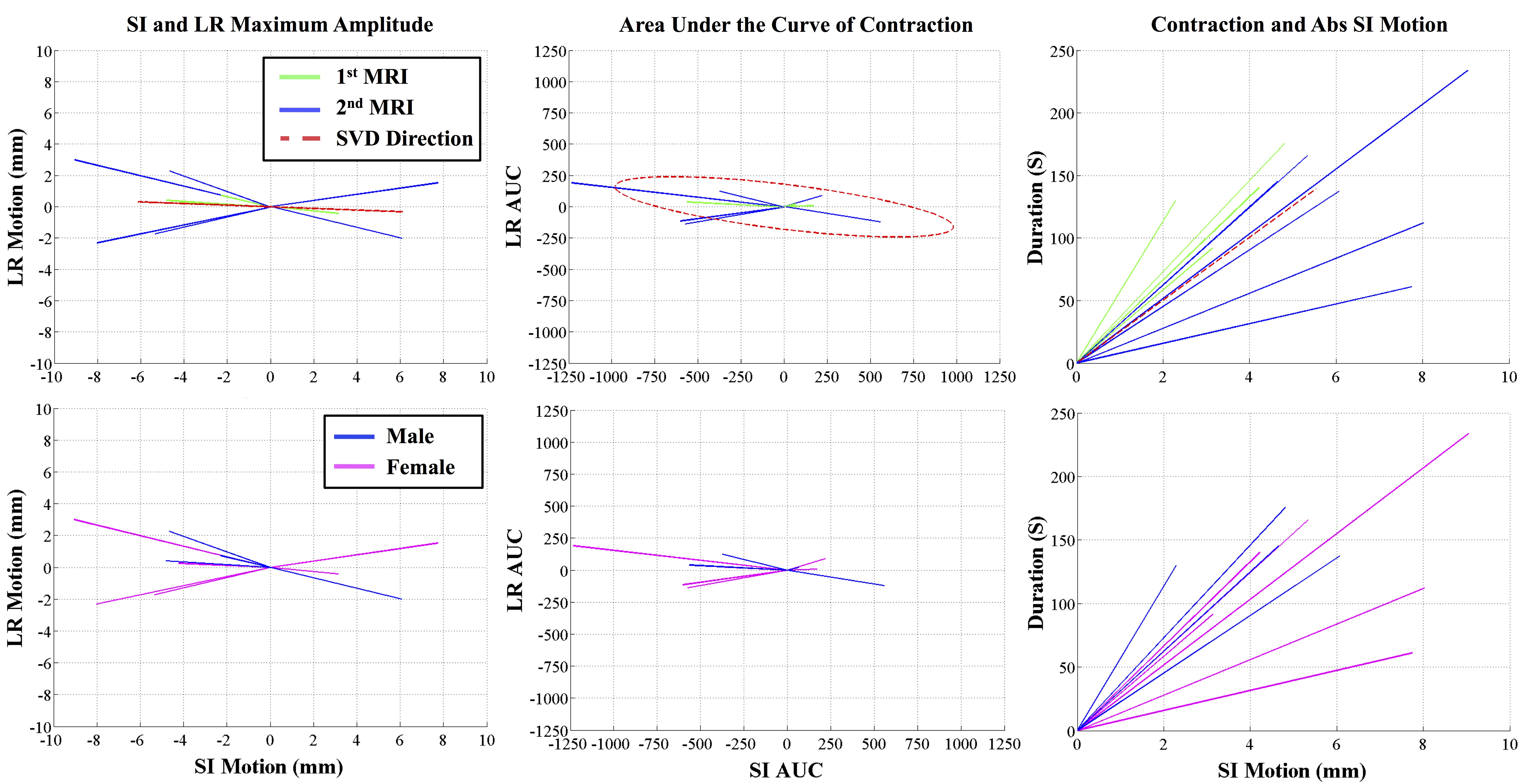
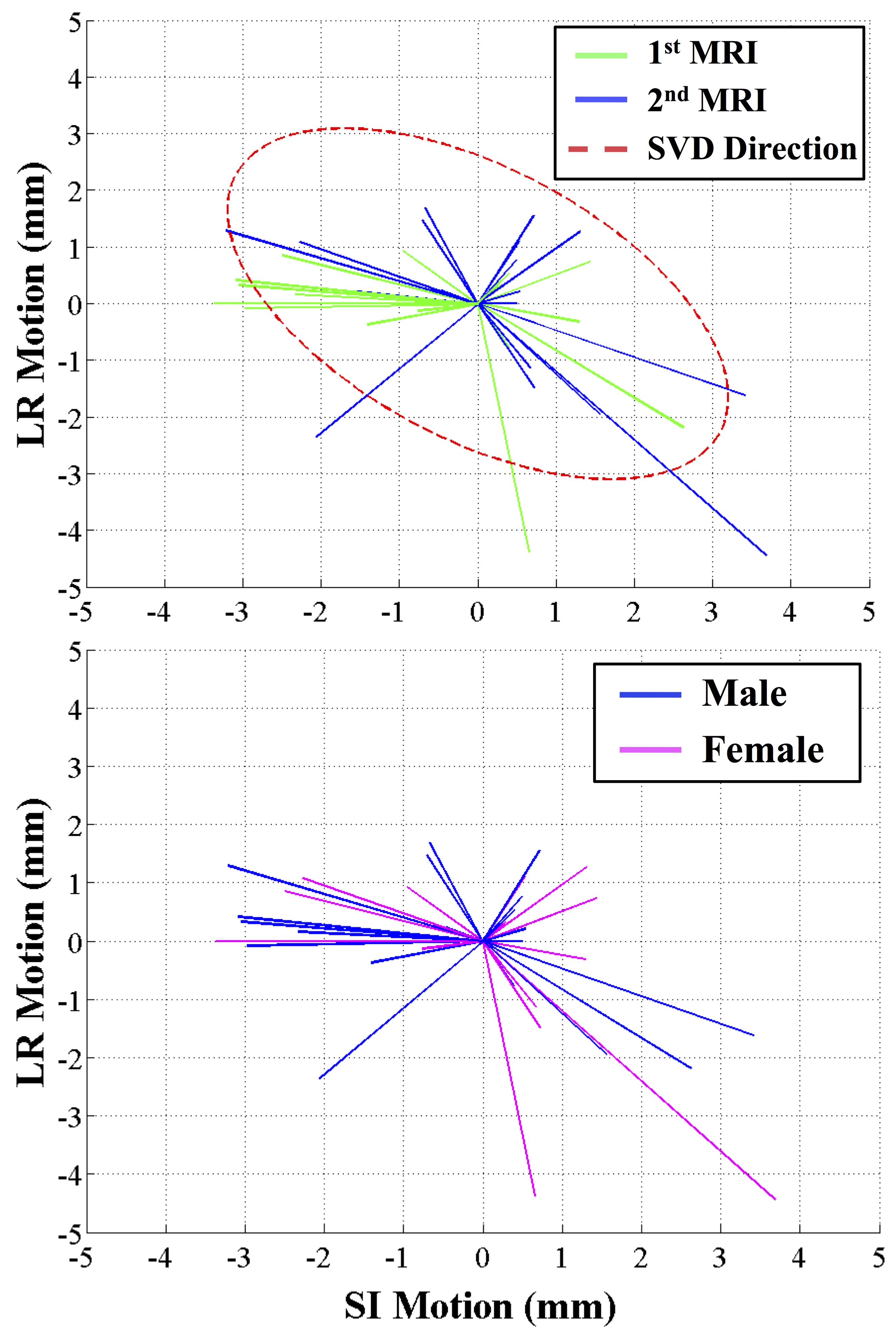
There were a total of 10 out of 52 scans that had contractions during the scan (4/26 during 1st MRI, 6/26 during 2nd MRI). The first row of figure 3 plots the differences in magnitude and direction, AUC, and duration of contractions between the first and second MRI. The magnitude of contraction motion and AUC is greater in later gestation compared to the 1st MRI scan. However the direction does not vary between gestation ages. The SVD (red ellipse) shows that magnitude and direction is dominant in the SI direction compared to LR motion. The mean duration of all the contractions is 145s. There were no fetal sex-specific differences in contraction motion observed. The mean amount of SI motion in contractions is 6 mm while LR is less than 1 mm. Figure 4 shows that the dominant direction of uterine motion in scans with out contractions (40 out of 52) is in the SI motion with a mean of 3 mm. There was no difference in bulk motion between MRI scans of each subject and related to fetal gender.Discussion and Conclusion
We have demonstrated that uterine and placental motion can be characterized using 3D golden angle radial GRE sequence with KWIC reconstruction and then using an image-based template-matching algorithm. Uterine contractions were observed in 4/26 scans and 6/26 scans in 14-16 weeks and 19-22 weeks, respectively. It was observed that uterine contraction is most significant in the SI direction and has mean amplitude of 6mm of uterine and placental motion. The direction, AUC, and duration of the scans are not affected by fetal gender and gestational age. This study can potentially further help develop proper scanning protocols for MRI studies with pregnant patients to avoid potential motion artifacts.Acknowledgements
Funding for project is provided by NICHD U01-HD087221.References
1. Coakley F V., Glenn OA, Qayyum A, Barkovich AJ, Goldstein R, Filly RA. Fetal MRI: A Developing Technique for the Developing Patient. Am J Roentgenol. 2004;182(1):243-252. doi:10.2214/ajr.182.1.1820243.
2. Prayer D, Brugger PC, Prayer L. Fetal MRI: Techniques and protocols. Pediatr Radiol. 2004;34(9):685-693. doi:10.1007/s00247-004-1246-0.
3. Hee KS, Dougherty L. Dynamic MRI with projection reconstruction and KWIC processing for simultaneous high spatial and temporal resolution. Magn Reson Med. 2004;52(4):815-824. doi:10.1002/mrm.20237.
4. Wu HH, Gurney PT, Hu BS, Nishimura DG, McConnell M V. Free-breathing multiphase whole-heart coronary MR angiography using image-based navigators and three-dimensional cones imaging. Magn Reson Med. 2013;69(4):1083-1093. doi:10.1002/mrm.24346.
Figures