4692
Evaluating Female Pelvic Pain with DISCO MRI: Pilot study in Pelvic Congestion and Uterine Fibroids1Radiology, University of California San Diego, La Jolla, CA, United States
Synopsis
Pelvic congestion and uterine fibroids are two common causes of female pelvic pain that can be treated with image guided vascular embolization and often evaluated with MRI prior to treatment. Vascular as well as anatomic imaging are important in evaluation and treatment planning for these two disease processes. Differential sampling with Cartesian ordering (DISCO)-MRI is a technique that allows for both high temporal and spatial resolution. This abstract demonstrates the utility of DISCO-MRI in evaluation and treatment planning for pelvic congestion and uterine fibroids.
Introduction
Chronic pelvic pain affects 15% of women 18 – 50 years old [1]. Pelvic congestion and uterine fibroids are two common causes of female pelvic pain that can be treated with image guided vascular embolization and often evaluated with MRI prior to treatment. Retrograde flow in the ovarian veins can often be seen in pelvic congestion, but requires rapid multiphasic imaging for visualization. If retrograde flow is appreciated in the ovarian veins in pelvic congestion, they can be embolized to help treat the symptoms. Uterine fibroid vascular supply usually emanates from branches of the uterine arteries, but can also be supplied from anomalous vessels. Planning for image guided uterine fibroid embolization may be guided by visualization of the feeding arterial supply and can be assessed with MRA. Evaluation of uterine fibroids and pelvic congestion could benefit from an MRI technique that allows for both high temporal and spatial resolution. DISCO (differential sampling with Cartesian ordering)-MRI [2] has demonstrated great utility in liver mass imaging, and also has potential for evaluation in pelvic MRI for uterine fibroids and pelvic congestion. This is the first study assessing the utility of DISCO-MRI in patients with pelvic congestion and uterine fibroids.Methods
A retrospective review was performed on the 15 most recent patients at our institute evaluated with MRI for uterine fibroids and pelvic congestion, including an acquisition with multiphasic dynamic contrast enhanced MRI (Gadobutrol) using DISCO -MRI at 3T. Of the 15 studies, 10 studies focused on fibroid evaluation and 5 studies focused on pelvic congestion. The DISCO protocol for fibroid evaluation weighted spatial resolution over temporal resolution with the following protocol: 13 phases, FOV 38 cm, slice thickness 2.8, 100 slices, flip angle 12, acceleration 1.8 phase x 2.2 slice, matrix 224 x 192, and BW 167. The DISCO protocol for pelvic congestion weighted temporal resolution over spatial resolution with the following protocol: 17 phases, FOV 38 cm, slice thickness 2.8, 100 slices, flip angle 12, acceleration 1.8 phase x 2.5 slice, matrix 192 x 160, and BW 167. In addition, conventional imaging was performed including coronal and axial T2 weighted single shot fast spin echo (FSE), sagittal and oblique axial T2 weighted fast relaxation FSE, and contrast enhanced liver acquisition with volume acceleration (LAVA-FLEX). A radiologist reviewed the 15 studies. In the uterine fibroid cases, the radiologist evaluated whether the uterine arteries were visible with DISCO and standard post contrast imaging. For the pelvic congestion cases, the radiologist determined if the patient had left renal vein reflux by evaluating the DISCO images and determining whether the left ovarian vein filled retrograde before the filling of the iliac veins.Results
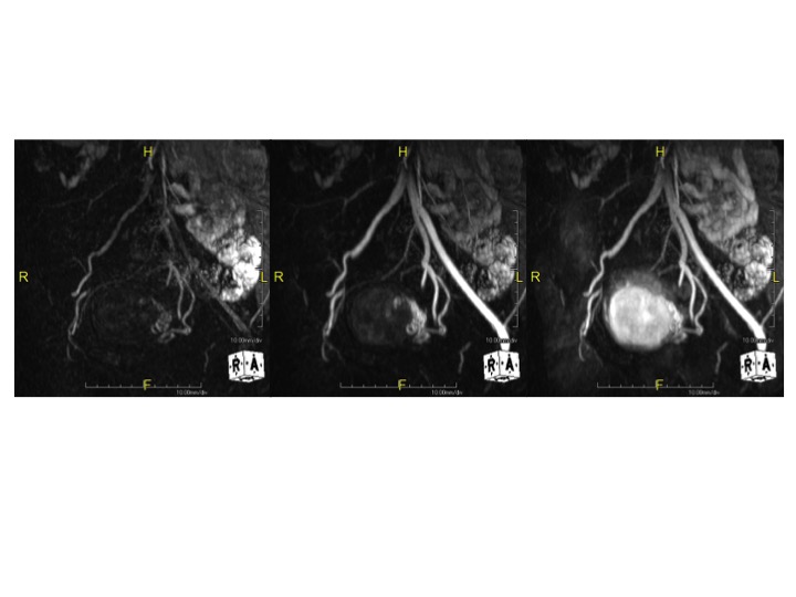
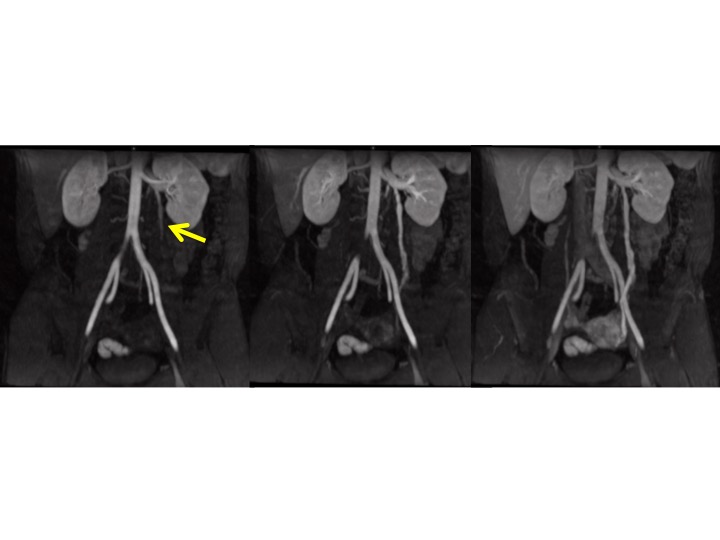
For the ten studies evaluating uterine fibroids, all 10 of them demonstrated clear visualization of the uterine arteries with DISCO and were not visible with standard post-contrast studies (Figure 1). Out of the five patients evaluated for pelvic congestion, four of the patients demonstrated left ovarian vein reflux prior to filling of the iliac veins with DISCO imaging (Figure 2). In these four patients, retrograde filling of the left ovarian vein was indiscernible with standard post contrast imaging. For one of the patients evaluated for pelvic congestion, no retrograde filling was appreciated nor were there dilated pelvic vessels on post contrast imaging (thus, pelvic congestion was not demonstrated by MRI).Discussion
Multiphasic contrast enhanced DISCO MRI can play a diagnostic role in evaluating female pelvic pain and planning for image guided procedures. The rapid capturing of multiple vascular flow phases, as demonstrated by this study, allows imaging diagnosis of reflux in the left ovarian vein which can be seen in pelvic congestion. In regards to uterine fibroid MRI, it is helpful to evaluate the feeding vessels of the fibroid and note anomalous vasculature prior to image guided embolization. For the 10 patients included in this study, early phase imaging with DISCO provided clear visualization of the uterine arteries without anomalous blood flood to the fibroids or uterus.Acknowledgements
GE HealthcareReferences
[1] Kuligowska E, Deeds L, Kang L. Pelvic Pain: Overlooked and Underdiagnosed Gynecologic Conditions. Radiographics: (25)1: 2005.
[2] Saranathan M, Rettman DW, Hargreaves BA, et al. Differential subsampling with Cartesian Ordering (DISCO): A High Spatio-temporal Resolution Dixon Imaging Sequence for Multiphasic Contrast Enhanced Abdominal Imaging. J Magn Reson Imaging. 35(6):2012.
Figures