4691
Investigation of correlation between IVIM and DCE-MRI on uterine cervical carcinoma1National Cancer Center/Cancer Hospital, Chinese Academy of Medical Sciences and Peking Union Medical College, Beijing, China, 2GE Healthcare Life Science, Beijing, China, 3GE Healthcare, MR Research China, Beijing, China
Synopsis
Perfusion was of great importance to access tumor properties. In this study, the potential correlations of perfusion parameters derived from IVIM (D*, f, and f D*) and DCE-MRI (Ktrans, Kep and Ve) for uterine cervical carcinoma (UCC) were investigated and were compared between pathological types. D* and f D* were positively correlated with Ktrans, Kep and Ve, respectively. Adenocarcinoma had higher f, Ktrans and Kep values than those of squamous cell carcinoma. Therefore, IVIM, as a non-invasive method, has potential to replace DCE-MRI to accurately access tumor perfusion properties, especially when perfusion differs among different pathological types of UCC.
Purpose
To investigate the correlation of IVIM and DCE-MRI derived perfusion parameters on different pathological types of uterine cervical carcinoma.Introduction
Dynamic contrast-enhanced magnetic resonance imaging (DCE-MRI) was recognized as a reliable method to evaluate tissue perfusion, while IVIM, as a non-invasive method, has also demonstrated microvessel density of tumor1-3. The correlation between the perfusion parameters derived from IVIM and DCE-MRI remained was barely studies. Moreover, the potential difference between different pathological types of uterine cervical carcinoma (UCC) has not been investigated using IVIM and DCE-MRI. The current study aimed to investigate the potential correlation of perfusion parameters between IVIM and DCE-MRI models and to explore the feasibility of applying IVIM on UCC, squamous cell carcinoma (SCC) and adenocarcinoma (AdC).Material and methods
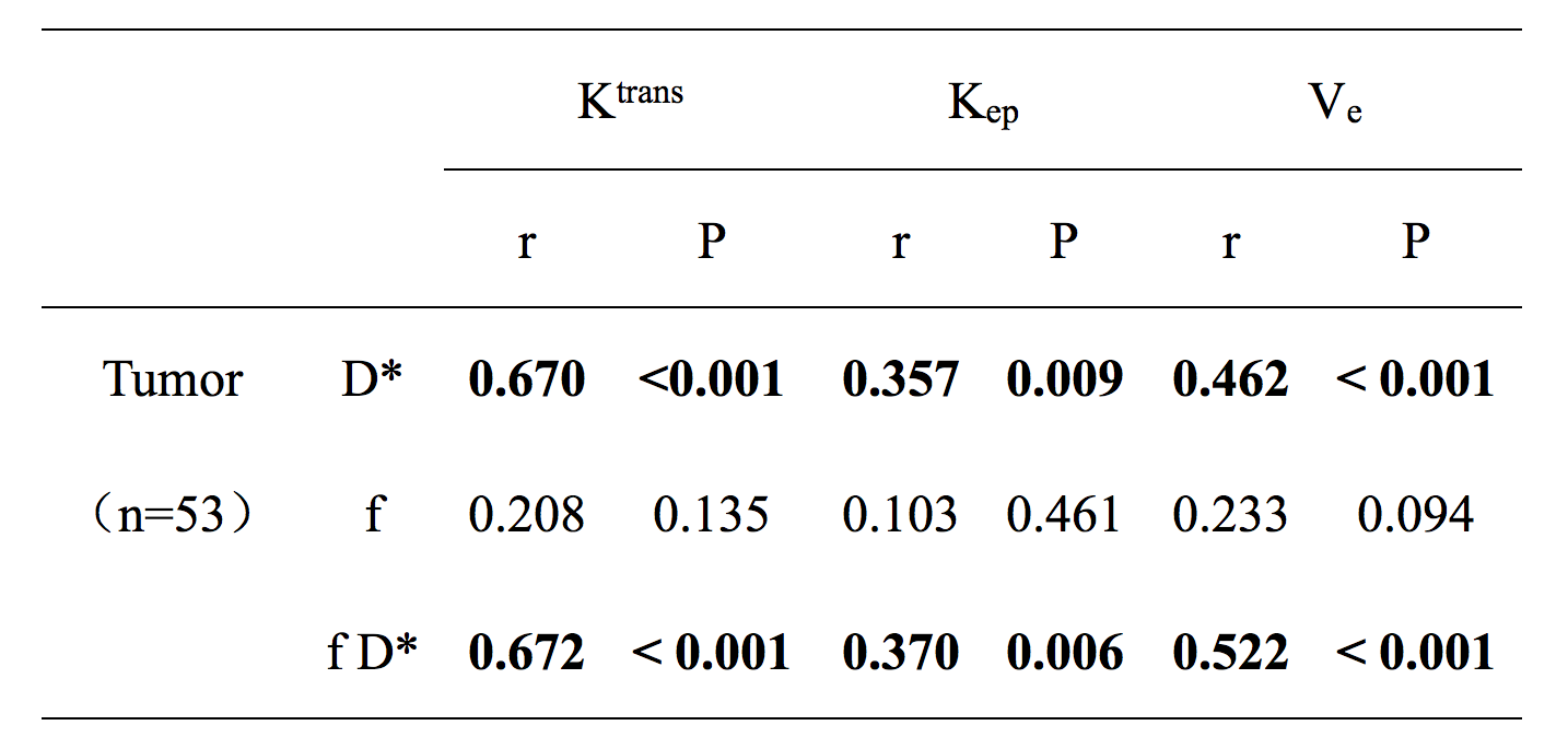
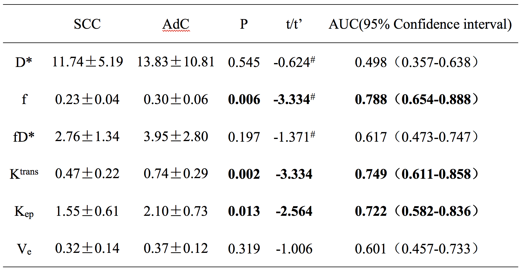
Fifty-three patients with newly diagnosed uterine cervical carcinoma were recruited in our hospital from May 2015 to October 2016. All of them undergone magnetic resonance imaging (MRI) examinations including both IVIM and DCE-MRI sequences prior to the treatment. Perfusion parameters including D*,f, and f D* (f multipled by D*) were derived from IVIM, and Ktrans, Kep and Ve were derived from DCE-MRI., The potential correlation between those two models on UCC, SCC and AdC groups were respectively analyzed using Spearman’s correlation. Independent sample t test and t’ test were conducted to compare the difference of those derived perfusion parameters between SCC and AdC groups. ROC was measured to compare the area under the curve (AUC) and analyze diagnostic efficiency and the cut-off of the most meaningful parameter.Results
D* and f D* were slightly to moderately positively correlated with all of the three parameters of DCE-MRI in UCC (r: 0.357-0.672), whereas f showed no correlation with any parameter of DCE-MRI (Table1). AdC were associated with higher f, Ktrans and Kep value than those of SCC (P: 0.002-0.013, Table2). Considering f value less than 0.29 as the threshold to diagnose AdC, the sensitivity, specificity and accuracy were calculated to be 63.64%, 92.86% and 86.79%, respectively.Discussion and conclusion
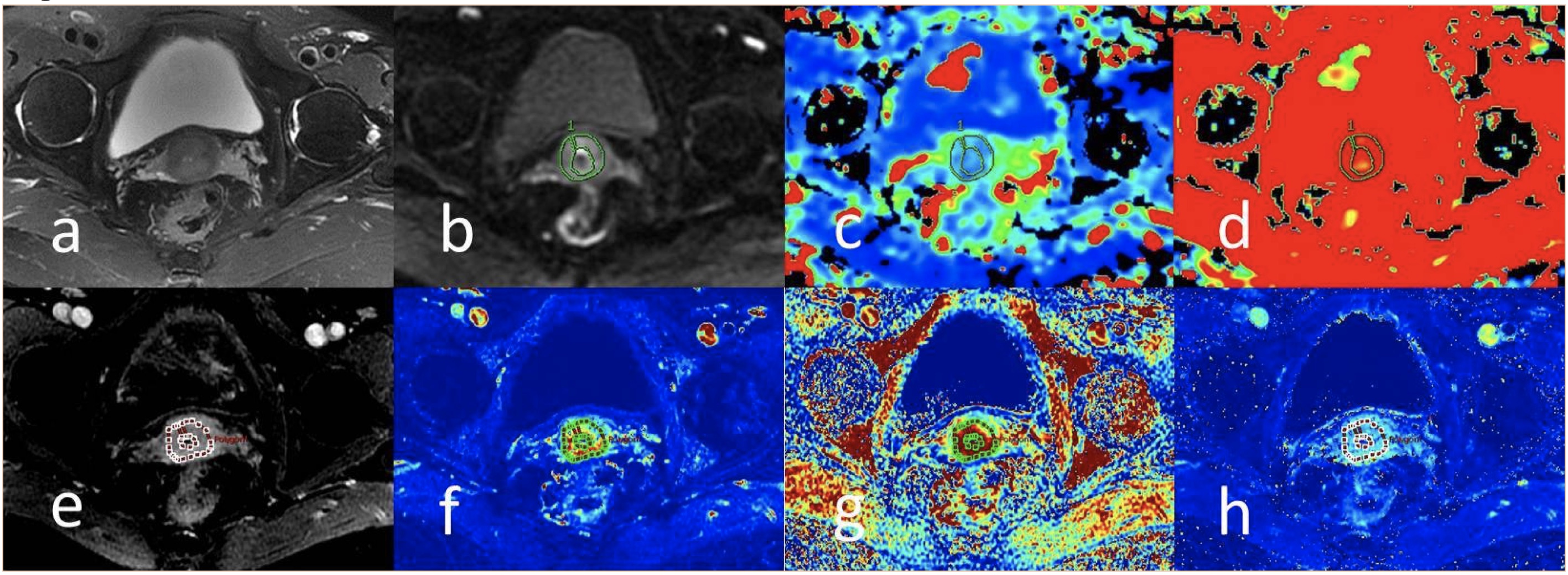
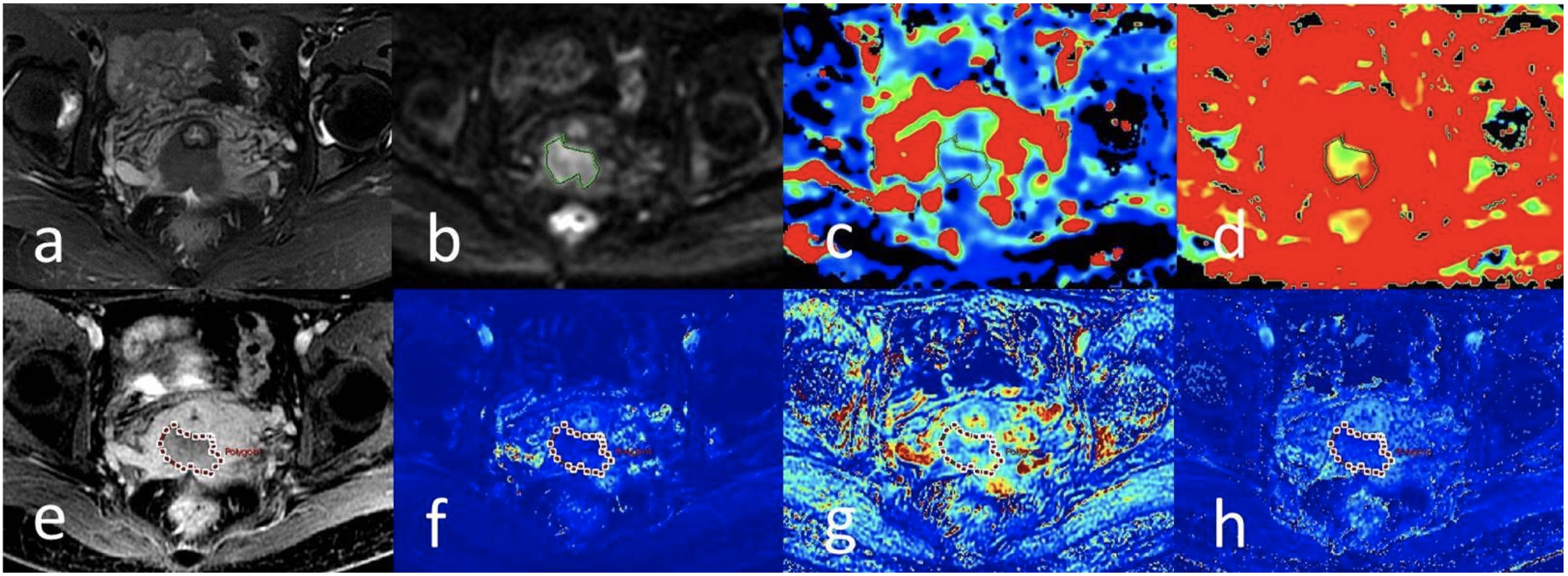
Typical cases of SCC and AdC were demonstrated in Figure1 and Figure 2, respectively. Consistent with the previous study1, fD* had slightly to moderate correlation with DCE-MRI. However, inconsistent with another study4, where f was observed to slightly to moderately correlate with Maxslop, CER and AUC of DCE-MRI, the reason may be due to different quantitative and semi-quantitative DCE-MRI models and different TE time for IVIM sequence. Positive correlations existed between perfusion parameters derived from IVIM and DCE-MRI in UCC, suggesting that IVIM could provide meaningful information on tumor perfusion. SCC usually demonstrates enhanced heterogeneously and tends to associate with necrotic change. On the other hand, the enhancement of AdC was often homogeneous and persistently. The variance of perfusion among different pathological types of UCC was observed as well, where IVIM was helpful to identify it non-invasively.Acknowledgements
1. Lee EY, Hui ES, Chan KK, et al. Relationship between intravoxel incoherent motion diffusion-weighted MRI and dynamic contrast-enhanced MRI in tissue perfusion of cervical cancers. Journal of magnetic resonance imaging : JMRI. Aug 2015;42(2):454-9.
2. Lee HJ, Rha SY, Chung YE, et al. Tumor perfusion-related parameter of diffusion-weighted magnetic resonance imaging: correlation with histological microvessel density. Magnetic resonance in medicine. Apr 2014;71(4):1554-8.
3. Cui Y, Zhang C, Li X, et al. Intravoxel Incoherent Motion Diffusion-weighted Magnetic Resonance Imaging for Monitoring the Early Response to ZD6474 from Nasopharyngeal Carcinoma in Nude Mouse. Scientific reports. Nov 17 2015;5:16389.
4. Zhou Y, Liu J, Liu C, et al. Intravoxel incoherent motion diffusion weighted MRI of cervical cancer - Correlated with tumor differentiation and perfusion. Magnetic resonance imaging. Oct 2016;34(8):1050-6.
References
No reference found.Figures