4688
Combining 2d RF excitation with multishot acquisition for reduced distortion imaging of the female pelvis.1Global MR Applications and Workflow, GE Healthcare, Boston, MA, United States, 2Global MR Applications and Workflow, GE Healthcare, Menlo Park, CA, United States, 3Clinical Development, GE Healthcare, Waukesha, WI, United States, 4Global MR Applications and Workflow, GE Healthcare, New York, NY, United States, 5Radiology Medical Research Lab, University of Arizona, Tucson, AZ, United States, 6Global MR Applications and Workflow, GE Healthcare, Houston, TX, United States
Synopsis
The combination of two-dimensional RF excitation with echo planar imaging has shown to be a useful technique for diffusion-weighted imaging of small anatomical features with high-resolution, such as in the pelvis or pancreas. However, compared to standard single shot dw-epi, the trade-off between resolution and matrix size becomes disadvantageous as the field of view increases. We propose to combine 2d RF excitation with multishot echo planar acquisition to enable high-resolution imaging with reduced distortion over larger FOV and present preliminary results obtained in the female pelvis.
Introduction
Diffusion weighted MRI (DW-MRI) has been increasingly used in the female pelvis as a non-invasive modality with the potential to provide quantitative information about suspected gynecologic malignancies [1]. A conventional diffusion imaging work-up typically relies on single-shot echo planar sequence (SS-EPI) with the addition of saturation bands as an attempt to reduce the FOV in the phase encoding direction and avoid fat contamination. Recently, a more efficient approach was proposed consisting of restricting the FOV using tailor-designed 2-dimensional spatial excitation pulses (rFOV EPI) [2]. A single-shot implementation of this technique is available on modern scanners and has been used routinely in pelvic and abdominal imaging [3]. However, the female pelvis remains a challenging anatomical area for rFOV SS-EPI due to the presence of strong susceptibilities. In this study, we propose to combine rFOV with multishot acquisition and present preliminary results.
Methods
A single spin echo pulse sequence was implemented which combines rFOV with multishot diffusion acquisition, along with a MUltishot Sensitivity Encoded (MUSE) algorithm to reconstruct the data acquired from multiple shots by jointly solving for phase-encoded data and motion-induced phase errors estimated for each shot [4]. All volunteers were consented according to the institutional review board at our institution. A human female volunteer was scanned on a 3T 70cm bore scanner (Signa Premier, GE Healthcare, USA) using the GEM anterior and posterior array. Diffusion images were collected Sagittally using a ss-EPI rFOV FOCUS sequence with TR/TE=4621/53.1ms full echo, Effective Echo Spacing (EESP)=0.7ms, 2mm slice thickness, 18cm square FOV, 92x92 acquisition matrix, 3 diffusion encoding applied respectively in x, y, z and b-values 50s/mm2 (NEX=2) and 600s/mm2 (NEX=12) with High Order Eddy Current Correction (HOECC), total scan time=3min19s; and an rFOV-enabled MUSE sequence with identical slice prescription and diffusion encoding scheme including HOECC, TR/TE=4500/54.8ms, EESP=0.35ms, 2shots, 92x92 acquisition matrix and total san time=6min27s.Results and Discussion
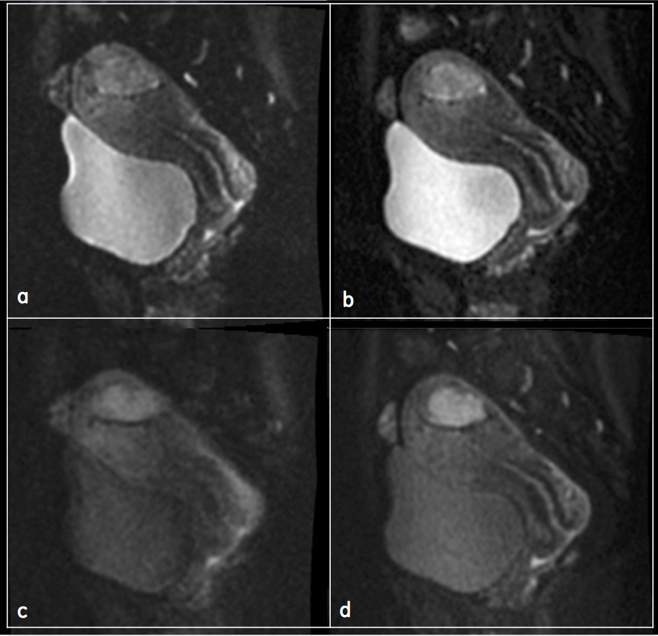
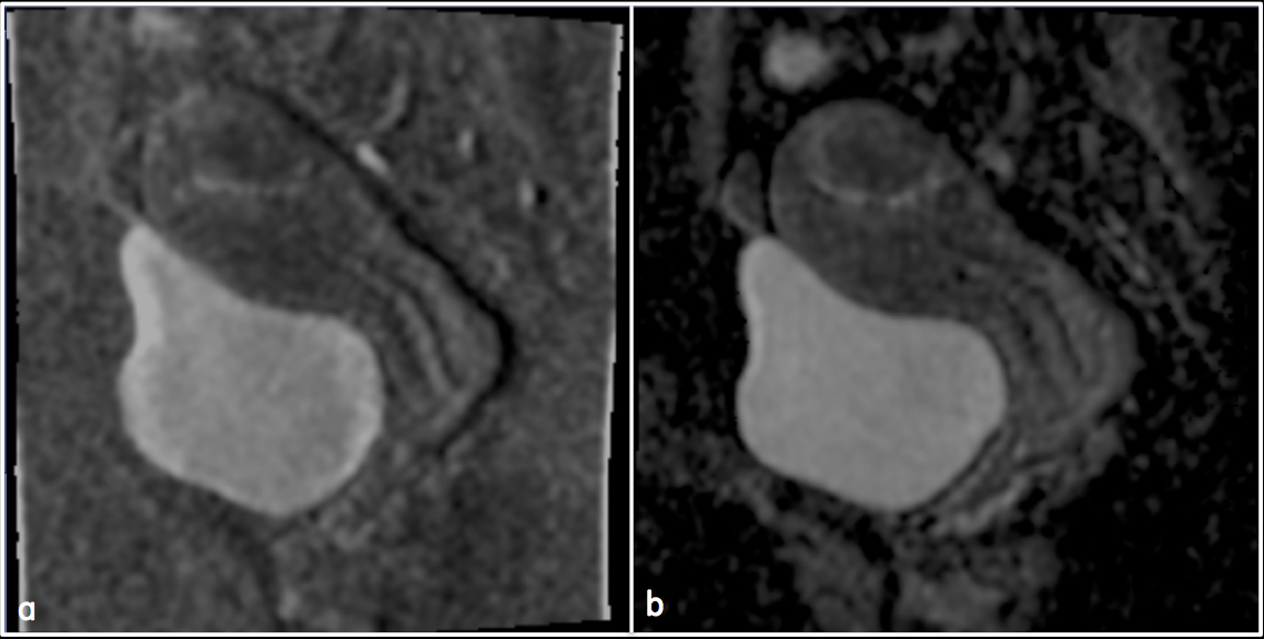
Figure 1 shows the rFOV SS-EPI and 2-shot rFOV MUSE images. Susceptibility artifacts are substantially reduced on the 2-shot rFOV MUSE images, where boundaries are sharper in both the low and high b-value images, such as around the rectal wall and cervical canal. Signal pile-up caused by strong off-resonances as observed along the anterior wall of the bladder on the low diffusion image (figure 1a) is also more severe in the rFOV SS-EPI compared to the 2-shot rFOV MUSE image. Apparent Diffusion Coefficient (ADC) maps for rFOV SS-EPI and 2-shot rFOV MUSE images are shown in Figure 2a and 2b respectively. The sharp contrast between organs of interest and surrounding tissue in the 2-shot rFOV MUSE image suggests that 2-shot rFOV MUSE is significantly more immune to physiological and systematic noise. The relative homogeneity of the ADC measurements throughout the bladder in the 2-shot rFOV MUSE image also serves as an indicator that the sequence is more robust. Efforts are currently underway to confirm these preliminary findings in a larger cohort.
Conclusion
Our initial results demonstrate that combing an rFOV excitation scheme with a multishot acquisition based on the MUSE reconstruction algorithm may be a viable option for large FOV, high SNR, high-resolution diffusion imaging of the female pelvis. It enables full coverage of the pelvic area without trading off the large matrix size needed for high spatial resolution, albeit at the expense of increased scan time. We anticipate that this protocol may prove useful not only for diagnosis but also for staging and monitoring of gynecological malignancies and are currently designing a study aimed at investigating the clinical practicality of this method.Acknowledgements
No acknowledgement found.References
- Whittaker, C.S., Coady, A., Culver, L., Rustin, G., Padwick, M., Padhani, A.R., 2009. Diffusion-weighted MR Imaging of Female Pelvic Tumors: A Pictorial Review. RG 29, 759–774. doi:10.1148/rg.293085130
- Saritas, E.U., Cunningham, C.H., Lee, J.H., Han, E.T., Nishimura, D.G., 2008. DWI of the spinal cord with reduced FOV single-shot EPI. Magn Reson Med 60, 468–473. doi:10.1002/mrm.21640
- Bhosale, P., Ma, J., Iyer, R., Ramalingam, P., Wei, W., Soliman, P., Frumovitz, M., Kundra, V., 2015. Feasibility of a reduced field-of-view diffusion-weighted (rFOV) sequence in assessment of myometrial invasion in patients with clinical FIGO stage I endometrial cancer. J. Magn. Reson. Imaging 43, 316–324. doi:10.1148/rg.323115736
- Chen, N.-K., Guidon, A., Chang, H.-C., Song, A.W., 2013. A robust multi-shot scan strategy for high-resolution diffusion weighted MRI enabled by multiplexed sensitivity-encoding (MUSE). NeuroImage 72, 41–47. doi:10.1016/j.neuroimage.2013.01.038
Figures