4671
PRAGMATIC ABILITIES IN MULTIPLE SCLEROSIS PATIENTS: AN RS-fMRI STUDY1Department of Advanced Biomedical Sciences, University of Naples "Federico II", Naples, Italy, 2University of Naples "Federico II", Naples, Italy, 3Institute of Biostructure and Bioimaging, National Research Council, Naples, Italy, 4Scuola Universitaria Superiore IUSS, Pavia, Italy, 55. IRCCS Fondazione Ospedale San Camillo, Venice, Italy
Synopsis
Multiple Sclerosis (MS) patients could experience communicative deficits in “pragmatics”, which is the ability to integrate context-dependent aspects of meaning beyond structural components of language. We evaluated relationships between pragmatics and functional connectivity (FC) of the bilateral inferior parietal lobule (the so-called Geschwind’s areas -GA-), in MS patients via a seed-based Resting-State fMRI analysis. We found a direct correlation between pragmatic scores and FC of both right (p=0.003) and left (p=0.009) GAs with the paracingulate cortex. Our results suggest that language is not only a left hemisphere function, and highlight a possible central role of paracingulate cortex in pragmatics.
Introduction
Cognitive functions have been largely investigated in multiple sclerosis (MS). Recent studies showed that MS patients could also experience communicative deficits, specifically in the so-called “pragmatics” (i.e. the ability to correctly integrate the context-dependent aspects of meaning beyond the structural components of language). While the classical model for language involves a posterior temporal region (Wernicke’s area) along with the inferior frontal gyrus (Broca’s area), several studies highlighted the role of the inferior parietal lobule (Geschwind’s area, GA) in high-level language processing, including comprehension of global coherence of narratives, global coherence of narratives, metaphor and irony comprehension as well as other pragmatic tasks. We performed a cross-sectional study to evaluate the relationship between pragmatic abilities and the functional connectivity (FC) of the bilateral GAs in MS patients.Methods
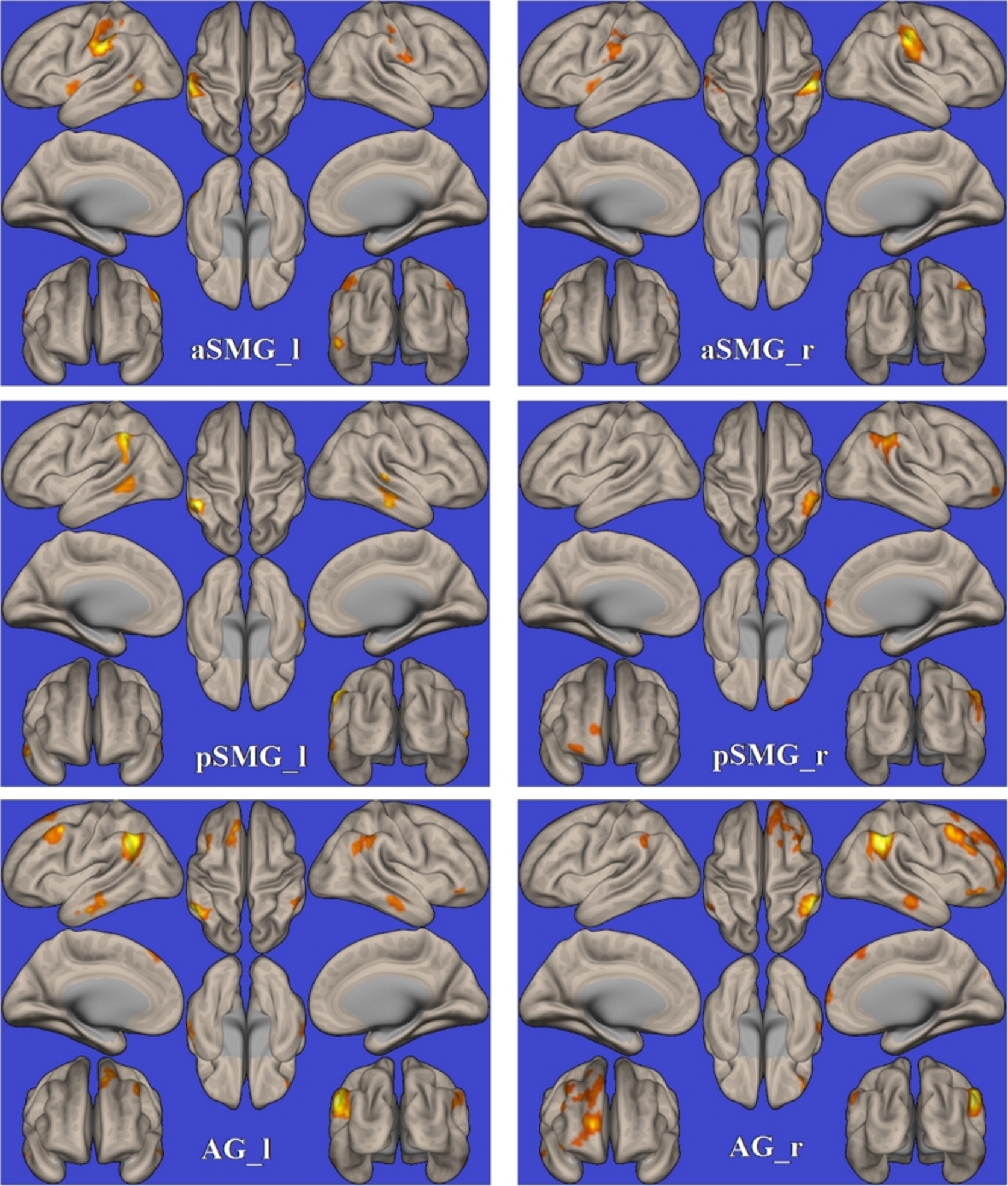
We enrolled 40 MS patients diagnosed according to the 2010 McDonalds criteria 1. Pragmatic abilities were assessed in all MS patients through a standardized test for the Italian speaking population (APACS, Assessment of Pragmatic Abilities and Cognitive Substrates) 2. MRI data were acquired on a 3 Tesla MR scanner (Trio, Siemens Medical Systems, Erlangen, Germany). Functional data were processed using the functional connectivity toolbox (CONN, v. 16.a, http://www.nitrc.org/projects/conn), which contains libraries for fMRI analysis based on the Statistical Parametric Mapping (SPM8) software package (http://www.fil.ion.ucl.ac.uk/spm). Briefly, pre-processing steps included the removal of the first five time points, to allow for instability of the initial MRI signal, leaving 195 time points, followed by the motion and slice timing correction, the temporal despiking by means of an hyperbolic tangent squashing function to limit outlier values, band-pass filtering (0.008 Hz<f<0.09 Hz), and spatial smoothing (using a 6-mm Full-Width at Half Maximum Gaussian kernel). Studies with a mean relative root-mean-square (RMS) of the translation parameters at each time point of 0.15 or higher (according to 3), or with more than 1.5mm displacement along or 1.5 degrees rotation around any of the three main axes, were discarded from the analysis. For each subject, BOLD signal time course was calculated over each subregion of both left and right GAs, namely the anterior and posterior division of the left (aSMG_l and pSMG_l) and right (aSMG_r and pSMG_r) Supramarginal Gyrus, and the right (AG_r) and left (AG_l) Angular Gyri as defined in the Harvard-Oxford atlas 4, and corresponding correlation maps of the BOLD signal across the brain were then generated for each patient (Figure 1). Correlations between the FC maps and the APACS Total scores were tested voxel-wise separately for each of the six selected seeds, including age, gender, RMS, and educational level in the model.Results
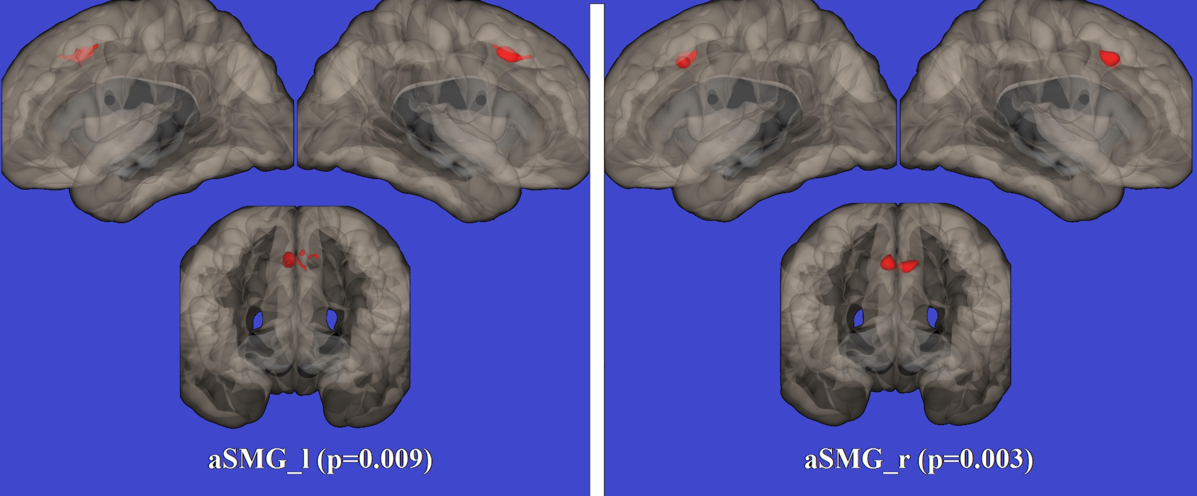
A cluster of significant direct correlation was observed between APACS scores and the FC of the right GA seed with the paracingulate cortex (p=0.003). A similar correlation between pragmatic abilities and paracingulate cortex was also proved when FC of the left GA seed was tested (p=0.009) (Figure 2).Discussion
To date, only a few studies have explored the role of the inferior parietal lobule in pragmatics, a field of great interest in neurodegenerative disorders due to the impact of communicative skills on patients’ social activities. Recent studies reported that MS patients could experience pragmatic deficits, similarly to other neurodegenerative disorders such as Parkinson and Alzheimer disease 5-7. However, little is known about the neuroanatomical functional network underpinning pragmatic skills as well as pragmatic impairment. Previous studies on high-level context-dependent language tasks, such as the comprehension of metaphors, irony 8-9, and indirect speech acts 10, pointed to both left and right GAs as a key cortical region, and led to models where the GA plays a fundamental role in supporting pragmatics and social communication 11. We found a significant correlation between the FC of bilateral GAs and the paracingulate cortex and pragmatic abilities, as assessed through the APACS Total scores. Importantly, the connection between the GA and the paracingulate cortex has a key role in stimulus-driven control of attention and diverting self-reflective thinking to salient external stimuli 12. Therefore, this network seems well placed for deriving the speakers’ meaning at the highest level of communication and for supporting appropriate pragmatic behavioral.Conclusion
We showed a direct correlation between FC of bilateral GAs and paracingulate cortex with pragmatic abilities in MS patients. Our results evidenced that language, when considered in the context-based communicative dimension, is not only a left hemisphere function, but it involves a more branched network over both hemispheres, which might be somehow disconnected in MS. Furthermore, our results highlight the possible central role of paracingulate cortex in pragmatic abilities.Acknowledgements
No acknowledgement found.References
1. Polman, C. H., Reingold, S. C., Banwell, B.,et al. (2011). Diagnostic criteria for multiple sclerosis: 2010 revisions to the McDonald criteria. Ann Neurol, 69(2), 292-302. doi:10.1002/ana.22366 2. Arcara, G., & Bambini, V. (2016). A Test for the Assessment of Pragmatic Abilities and Cognitive Substrates (APACS): Normative Data and Psychometric Properties. Front Psychol, 7, 70. doi:10.3389/fpsyg.2016.00070 3. Van Dijk, K. R., Sabuncu, M. R., & Buckner, R. L. (2012). The influence of head motion on intrinsic functional connectivity MRI. Neuroimage, 59(1), 431-438. doi:10.1016/j.neuroimage.2011.07.044 4. Desikan, R. S., Ségonne, F., Fischl, B., et al. (2006). An automated labeling system for subdividing the human cerebral cortex on MRI scans into gyral based regions of interest. Neuroimage, 31(3), 968-980. 5. Carotenuto, A., Arcara, G., Orefice, G., et al. (2017). Communication in Multiple Sclerosis: Pragmatic Deficit and its Relation with Cognition and Social Cognition. Arch Clin Neuropsychol, 1-12. doi:10.1093/arclin/acx061 6. Cuerva, A. G., Sabe, L., Kuzis, G., et al. (2001). Theory of mind and pragmatic abilities in dementia. Neuropsychiatry Neuropsychol Behav Neurol, 14(3), 153-158. 7. McNamara, P., & Durso, R. (2003). Pragmatic communication skills in patients with Parkinson's disease. Brain Lang, 84(3), 414-423. 8. Bambini, V., Gentili, C., Ricciardi, E., et al. (2011). Decomposing metaphor processing at the cognitive and neural level through functional magnetic resonance imaging. Brain Res Bull, 86(3-4), 203-216. doi:10.1016/j.brainresbull.2011.07.015 9. Spotorno, N., Koun, E., Prado, J., et al. (2012). Neural evidence that utterance-processing entails mentalizing: the case of irony. Neuroimage, 63(1), 25-39. doi:10.1016/j.neuroimage.2012.06.046 10. Basnakova, J., Weber, K., Petersson, K., et al. (2014). Beyond the language given: the neural correlates of inferring speaker meaning. Cereb Cortex, 24(10), 2572-2578. doi:10.1093/cercor/bht112 11. Catani, M. Bambini, V. A model for Social Communication And Language Evolution and Development (SCALED). Curr Opin Neurobiol. 2014 Oct;28:165-71. doi: 10.1016/j.conb.2014.07.018. Epub 2014 Aug 25. 12. Bays, P. M., Singh-Curry, V., Gorgoraptis, N., et al. Integration of goal- and stimulus-related visual signals revealed by damage to human parietal cortex. J Neurosci, 30(17), 5968-5978. doi:10.1523/JNEUROSCI.0997-10.2010Figures