4647
The vascular fingerprint of non-balanced BOLD SSFP coherence pathways: gradient echoes show spin echo behavior1MPI for biological Cybernetics, Tuebingen, Germany, 2Biomedical Magnetic Resonance, University of Tuebingen, Tuebingen, Germany
Synopsis
The S2-SSFP echo (the echo before the RF pulse in non-balanced SSFP sequences, or F-0, PSIF, CE-FAST, T2-FFE) is a refocused echo and has thus been proposed for BOLD imaging to suppress larger vessels, similar to the classical spin echo. Here we demonstrate in Monte Carlo simulations that the primary S1-SSFP gradient echo shows nearly identical properties as the S2-SSFP echo. Both echoes of this non-balanced SSFP sequence can be used for BOLD imaging exhibiting a pure spin echo behavior, i.e suppression of larger vessels.
Introduction
The vessel size dependence of the BOLD effect is often divided into two regimes: the diffusion narrowing regime for small vessel sizes of up to about 5-10 mm radius, and the static dephasing regime for larger vessels1-4. In the static dephasing regime, dephasing effects around vessels are almost completely refocused for spin echoes, but remain for gradient echoes and thus making them sensitive to larger vessels. Therefore, it is generally accepted that spin echoes are more sensitive to oxygenation changes located within micro vessels and thus might be closer to the neuronal event compared to gradient echoes. As an alternative to spin and gradient echoes, balanced SSFP and S2-SSFP (the non-balanced SSFP echo before the RF pulse) have been proposed and analyzed for BOLD imaging5-7,9. In this contribution we present an analysis of the BOLD sensitivity of non-balanced SSFP as a function of vessel size for several coherence pathways ranging from F-2 to F2, including the F+0 “gradient echo”, the F-0 “S2-echo” and higher order coherences.Methods
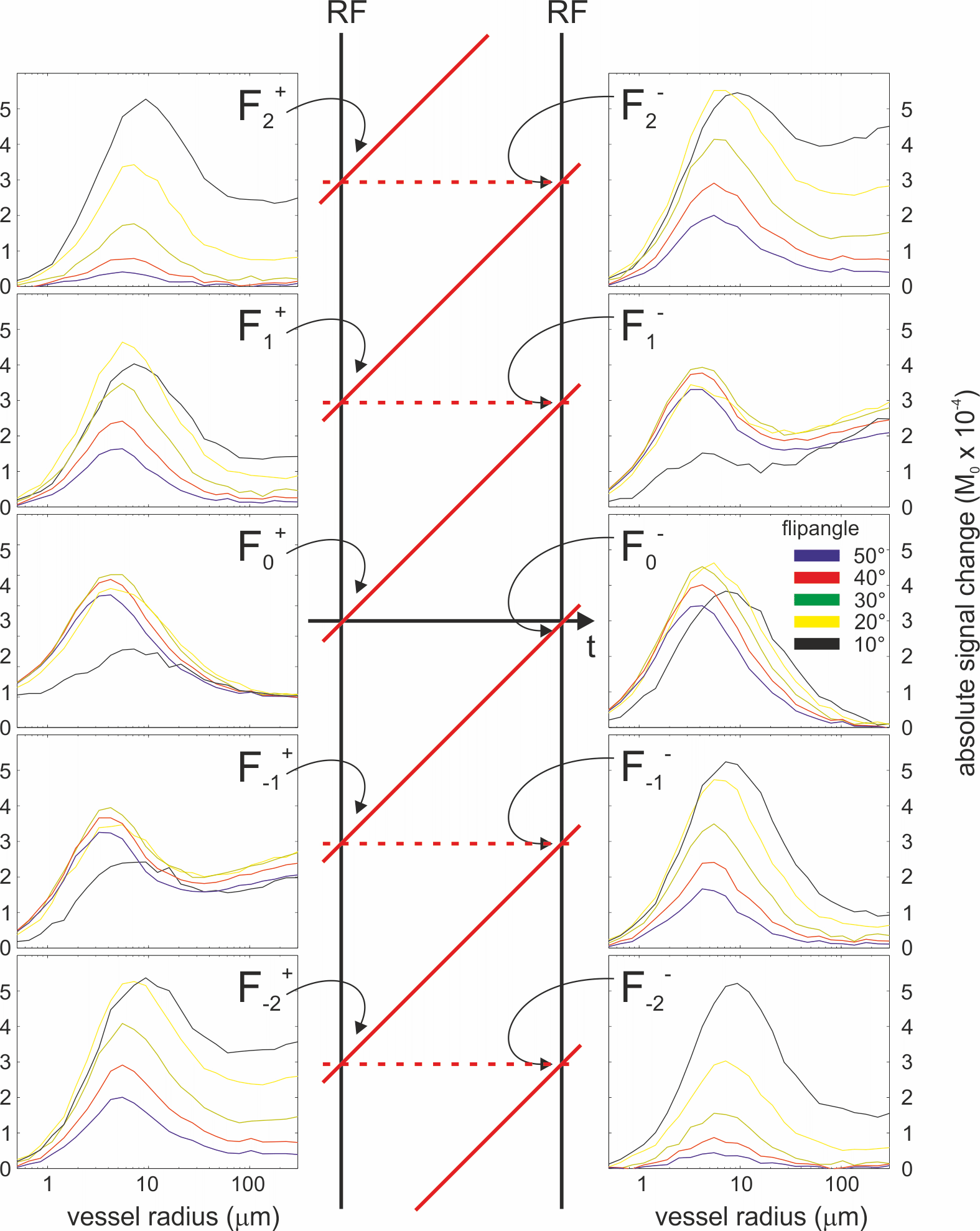
The MR signal formation of the extravascular water proton magnetization exposed to an extended train of RF pulses during their random walk through the neurovascular network was modeled for artificial cylinders with different diameter and random orientation to B0 according to1,7. For simplicity, only the resting state using Y = 80% (Δχ of 0.022 ppm) was compared to the activated, fully oxygenated state with Y = 100%. The BOLD signal, i.e. the signal difference between activated and resting state were calculated as absolute signal differences in units of M0, which corresponds to the contrast-to-noise of the BOLD effect. In all simulations, the steady state balanced SSFP signal profile was calculated at 32 different phase offsets between ± π, for different flip angles between 10° and 50°, TR=10ms, and different field strength from 3T to 9T, and a blood (cylinder) volume of 2%. Extravascular relaxation times as a function of B0 were calculated as T1= 1/(0.003*B0*B0 - 0.0791*B0 + 0.9247) and T2= 1/(1.74*B0 + 7.77) according to8,9. The resulting bSSFP signal profile after and before the RF pulse was then Fourier transformed to derive 32 different non-balanced SSFP coherence pathways ranging from F+-15 to F+16 (after RF pulse) and F--15 to F-16 (before RF pulse).Results
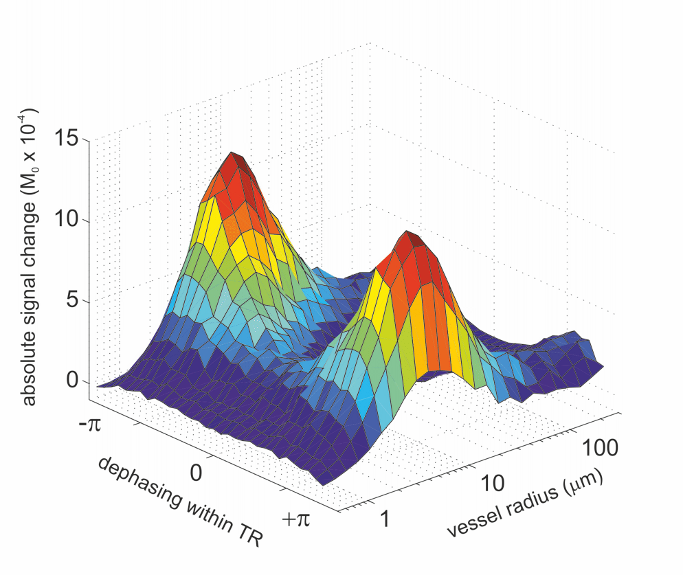
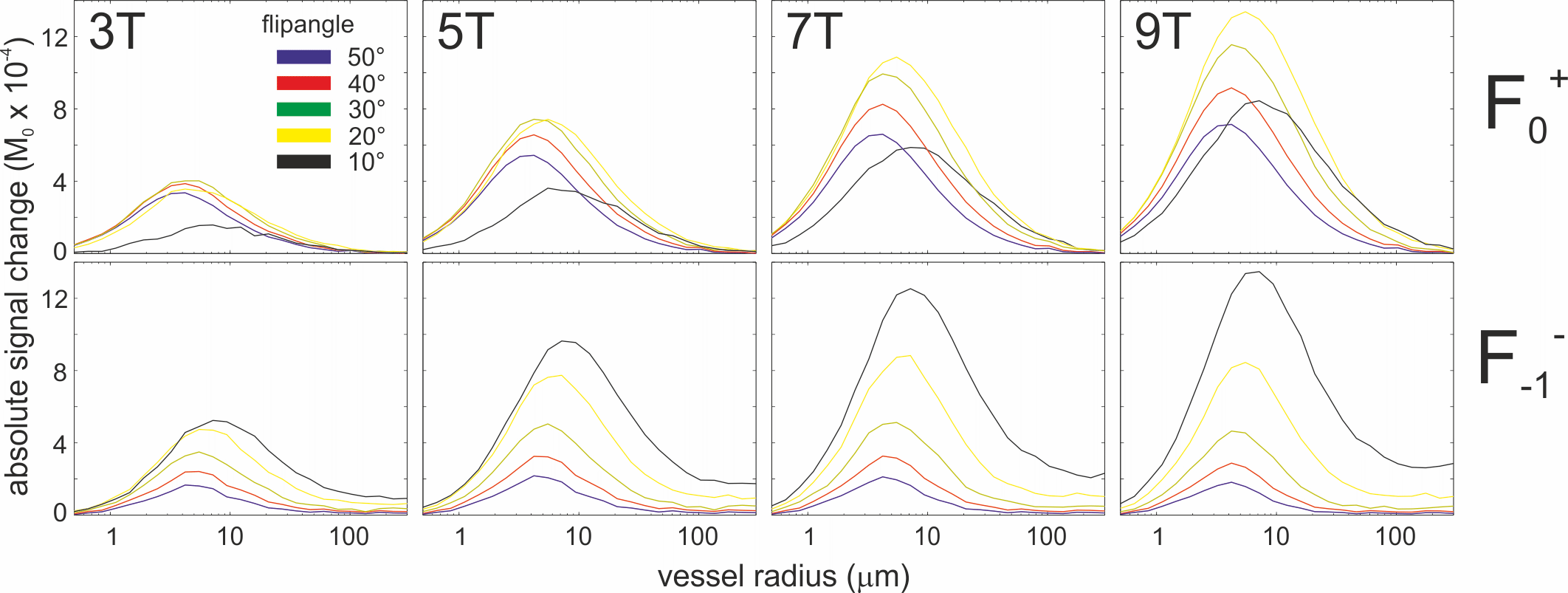
Fig. 1 shows an example of the simulated bSSFP profile (centered between RF pulses, 3T, 30° flip angle, TR=10ms, extravascular T1=1400ms and T2=77ms) between ± π and for cylinder radii ranging from 0.5 μm to 300 μm. The reported spin echo behavior (i.e. small BOLD effect for large vessels) is visible across the entire bSSFP profile, however, the BOLD signal change gets significantly larger at about ±0.7 π dephasing close to the stop bands. The profiles shown in Fig. 2 have been calculated as coefficients of the Fourier transformed bSSFP profile and represent the BOLD sensitivity for different echoes or pathways after (+) and before (-) the RF pulse. The F-0 path or S2-SSFP has been used for fMRI in several studies5,6, and it shows the expected spin echo property as it is a (partially) refocused echo path. Interestingly, the F+0 “gradient echo” basically shows the same spin echo-like behavior, i.e. a vanishing BOLD signal change for larger vessels. All other higher-order echo pathways exhibit an increasingly stronger gradient echo profile (although flip angle dependent) with increasingly larger contributions from larger vessels. Fig. 3 compares the BOLD signal change for F+0 and F--1 as a function of field strength B0. Both plots show an about linear increase with field strength, assuming identical M0 for all field strengths. Assuming a scaling10 power of 1.7 for M0 with B0 the BOLD signal change at 9T is about 20 times stronger than at 3T.Discussion
There is no need to use the refocused S2-SSFP echo to achieve a spin echo-like vessel size sensitivity for BOLD fMRI. The primary S1-SSFP echo shows nearly identical properties and is thus equally well suited as “spin echo” BOLD sequence. The spin echo behavior of this steady state gradient echo F+0 (FISP, FAST, GRASS, FFE, S1-SSFP, which is not identical to the T1-weighted RF-spoiled gradient echo) and for the F-0 echo (PSIF, CE-FAST, T2-FFE, S2-SSFP) was already theoretically predicted for static dephasing within a Lorentzian frequency distribution by Ganter11. This regime is similar to the quasi static dephasing regime for vessel radii larger than about 50 μm, as the diffusion length within TR is much smaller than the large vessel size.Acknowledgements
This work was supported by DFG SCHE658/12 Koselleck and the Max Planck SocietyReferences
1. Weisskoff, R., et al., 1994. Microscopic susceptibility variation and transverse relaxation: Theory and experiment. Magn. Reson. Med. 31, 601–610.
2. Boxerman, J. L., Bandettini, P. A., Kwong, K. K., Baker, J. R., Davis, T. L., Rosen, B. R., & Weisskoff, R. M. (1995). The intravascular contribution to fMRI signal change: Monte Carlo modeling and diffusion-weighted studies in vivo. Magnetic Resonance in Medicine, 34(1), 4–10.
3. Yablonskiy DA, Haacke EM. Theory of NMR signal behavior in magnetically inhomogeneous tissues: the static dephasing regime. Magn Reson Med. 1994 Dec;32(6):749-63.
4. Kiselev VG, Posse S. Analytical model of susceptibility-induced MR signal dephasing: effect of diffusion in a microvascular network. Magn Reson Med. 1999 Mar;41(3):499-509.
5. Barth, M., Meyer, H., Kannengiesser, S. A. R., Polimeni, J. R., Wald, L. L., & Norris, D. G. (2010). T2 weighted 3D fMRI using S2-SSFP at 7 tesla. Magnetic Resonance in Medicine, 63(4), 1015–1020.
6. Goa PE, Koopmans PJ, Poser BA, Barth M, Norris DG. BOLD fMRI signal characteristics of S1- and S2-SSFP at 7 Tesla. Front Neurosci. 2014 Mar 13;8:49. doi: 10.3389/fnins.2014.00049. eCollection 2014.
7. Báez-Yánez MG, Ehses P, Mirkes C, Tsai PS, Kleinfeld D, Scheffler K. The impact of vessel size, orientation and intravascular contribution on the neurovascular fingerprint of BOLD bSSFP fMRI. Neuroimage. 2017 Sep 8;163:13-23. doi: 10.1016/j.neuroimage.2017.09.015.
8. Uludağ K, Müller-Bierl B, Uğurbil K. An integrative model for neuronal activity-induced signal changes for gradient and spin echo functional imaging. Neuroimage 2009;48:150–65.
9. Khajehim M, Nasiraei Moghaddam A. Investigating the spatial specificity of S2-SSFP fMRI: A Monte Carlo simulation approach. Magn Reson Imaging. 2017 Apr;37:282-289. doi: 10.1016/j.mri.2016.11.016. Epub 2016 Nov 24.
10. Pohmann R, Speck O, Scheffler K. Signal-to-noise ratio and MR tissue parameters in human brain imaging at 3, 7, and 9.4 tesla using current receive coil arrays. Magn Reson Med. 2016 Feb;75(2):801-9. doi: 10.1002/mrm.25677. Epub 2015 Mar 29.
11. Ganter C. Static susceptibility effects in balanced SSFP sequences. Magn Reson Med. 2006 Sep;56(3):687-91.
Figures