4645
Caffeine enhances BOLD response to electrical whisker pad stimulation in rats1Center for Advanced Molecular Imaging and Translation, Chang Gung Memorial Hospital, Taoyuan, Taiwan, 2Department of Biomedical Imaging and Radiological Science, China Medical University, Taichung, Taiwan
Synopsis
The effects of caffeine on BOLD responses are still controversial. The potential explanation to this discrepancy may be associated with different dietary caffeine consumption among subjects. Here, we test the effect of caffeine on BOLD responses by using the caffeine-naïve animals. By reducing the baseline cerebral blood flow and therefore increasing the baseline deoxygenation concentration, caffeine significantly enhanced the magnitude of BOLD responses to the electrical whisker pad stimulation. These findings may suggest that caffeine-naïve animals could be a potential model to shed more light on effects of caffeine on BOLD fMRI measurements.
Introduction:
Caffeine is one of the most widely popular neurostimulant in the world, and it has various pharmacologic effects on the brain. The most well-known function of caffeine is that it acts as a nonselective antagonist of adenosine receptors, which causes the constriction of the blood vessels and reduces the cerebral blood flow (CBF), leading to the increases in deoxyhemoglobin concentration in the baseline condition. Thus, studies have been shown that caffeine has the ability to boost the blood oxygen level-dependent (BOLD) response during the active condition1, 2. However, there still exist reports that are controversial3, 4. The potential explanation to this discrepancy may be associated with different dietary caffeine consumption among human subjects5. The animal model, treated as the caffeine-naïve subjects, may has its potential role in understanding the impact of caffeine ingestion on brain physiology. Therefore, a central goal of this study is to exam whether we can mediate the BOLD response in rats by using caffeine even under anesthesia condition.Methods:
Study design: All animal experiments were approved by the local Institutional Animal Care and Use Committee. Seven Sprague-Dawley (SD) rats (260-340g) were scanned by using a 7T animal MRI scanner (Bruker ClinScan 70/30) under alpha-chloralose (80mg/kg) anesthesia. To map the primary somatosensory cortex barrel field (S1BF), needle electrodes were inserted under the skin of the left whisker pad. A stimulator supplied current of 3.5 mA and stimulation frequency of 3Hz. The electrical stimulation paradigm was OFF-ON-OFF-ON- OFF-ON-OFF-ON-OFF-ON-OFF, where OFF=75 seconds and ON=15 seconds. MRI measurement: The BOLD fMRI images were acquired before and after caffeine (40mg/kg) injection with the following parameters: TR=1000 ms, TE=25 ms, flip angle=90°, FOV=30 mm, and matrix size=64×64. For the post-caffeine section, images were obtained 40 minutes after the i.v. injection. Evaluation of physiological parameter: Mean arterial blood pressure (MABP) was measured in a separate group of 3 animals under the same experimental conditions but outside the scanning room. The purpose of this experiment was to test the effect of electrical stimulation on the physiological condition under caffeine.Results and discussion:
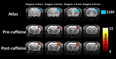
Caffeine effect on baseline BOLD signal: The baseline BOLD signals before and after caffeine injection are 321 ± 43 and 293 ± 48, respectively. The baseline BOLD signal significantly decreased in the caffeine condition (P=0.03), suggesting a higher level of deoxyhemoglobin in this baseline state. The magnitude of BOLD response straightforwardly relies on the difference in BOLD signals between rest and activation status. Both lower baseline signal and intensive stimulation can benefit the magnitude of BOLD response. Thus, it is expected to increase the subsequent BOLD response to the electrical stimulation. BOLD responses to stimulation with caffeine injection: Shown in Fig. 1 are maps of BOLD response induced by left whisker pad stimulation before and followed 40 min later by caffeine injection. Before the caffeine injection, 3.5mA whisker pad stimulation evoked the significantly positive BOLD response in the contralateral S1BF. Following caffeine, caffeine enhanced the magnitude of BOLD response in contralateral S1BF. The average percentage signal change of the BOLD response in contralateral S1BF shows a significant increase from 3.22 ± 0.29% to 7.14 ± 1.23% (Fig 2a, P<0.05). Quantifications of t-values are plotted in Fig. 2b. It is clear that t-values exhibited a significant improvement in statistical power in S1BF after caffeine injection (P = 0.05). Impact of caffeine on blood pressure: Caffeine injection itself did not alter the MABP during the whisker pad stimulation. The MABP before and after caffeine were respectively 106.9 ± 15.2 mmHg and 105.9 ± 15.8 mmHg (P=0.38), suggesting the BOLD responses to electrical stimulation under caffeine without the confounding effects of MABP changes.Conclusion
In this study, we demonstrate that with the pharmacological modulation of caffeine, the augmentation of BOLD response could be observed in the animals even under anesthesia condition for the first time. In the current study, the fixed caffeine dosage of 40 mg/kg through an injection was used in rats. With the fixed dosage, the metabolic rates of caffeine may not bias results. Meanwhile, the caffeine injection can occur without repositioning the animals. The same levels of magnetic field homogeneity and shimming make the direct comparison between pre- and post-caffeine sessions easier. All these findings may suggest that caffeine-naïve animals could be a potential model to shed more light on effects of caffeine on BOLD fMRI measurements.Acknowledgements
No acknowledgement found.References
1. Mulderink TA, Gitelman DR, Mesulam MM, Parrish TB. On the use of caffeine as a contrast booster for BOLD fMRI studies. NeuroImage 2002; 15: 37-44.
2. Chen Y, Parrish TB. Caffeine's effects on cerebrovascular reactivity and coupling between cerebral blood flow and oxygen metabolism. NeuroImage 2009; 44: 647-52.
3. Liau J, Perthen JE, Liu TT. Caffeine reduces the activation extent and contrast-to-noise ratio of the functional cerebral blood flow response but not the BOLD response. NeuroImage 2008; 42: 296-305.
4. Liu TT, Behzadi Y, Restom K, Uludag K, Lu K, Buracas GT et al. Caffeine alters the temporal dynamics of the visual BOLD response. NeuroImage 2004; 23: 1402-13.
5. Laurienti PJ, Field AS, Burdette JH, Maldjian JA, Yen YF, Moody DM. Dietary caffeine consumption modulates fMRI measures. NeuroImage 2002; 17: 751-7.

