4639
Demonstration of brain tumor-induced neurovascular uncoupling within the language network at ultra-high fieldShruti Agarwal1, Jun Hua2,3, Haris I. Sair1, Hanzhang Lu2,3, and Jay J. Pillai1,4
1Division of Neuroradiology, Russell H. Morgan Department of Radiology and Radiological Science, Johns Hopkins University School of Medicine, Baltimore, MD, United States, 2Division of MR Research, Russell H. Morgan Department of Radiology and Radiological Science, Johns Hopkins University School of Medicine, Baltimore, MD, United States, 3F. M. Kirby Research Center For Functional Brain Imaging, Kennedy Krieger Institute, Baltimore, MD, United States, 4Department of Neurosurgery, Johns Hopkins University School of Medicine, Baltimore, MD, United States
Synopsis
False-negative activations caused by neurovascular uncoupling (NVU) can lead to erroneous interpretation of clinical BOLD fMRI examinations. At 7T, spatial specificity can be improved relative to clinical 3T imaging. In this study, we demonstrate that NVU within the language network may affect the resting-state (rsfMRI) frequency domain metric ALFF (amplitude of low-frequency fluctuation) and breathhold cerebrovascular reactivity (BH CVR) maps as evident in the criterion standard task fMRI at ultra-high field despite known substantial BOLD signal-to-noise ratio advantages provided by higher field strength, which may not fully mitigate the effects of such NVU.
Purpose
False-negative activations caused by neurovascular uncoupling (NVU) can lead to erroneous interpretation of clinical BOLD fMRI examinations. At ultrahigh field (7 Tesla), spatial specificity can be improved relative to standard 3T imaging.1,2 Brain tumor-related NVU has been demonstrated at 7T within the sensorimotor network.3 The purpose of this study is to demonstrate that NVU within the language network may affect the resting-state (rsfMRI) frequency domain metric ALFF (amplitude of low-frequency fluctuation) and breath-hold cerebrovascular reactivity (BH CVR) maps as evident in the criterion standard task fMRI at ultra-high field despite known substantial BOLD signal-to-noise ratio advantages provided by higher field strength, which may not fully mitigate the effects of such NVU. Resting-state frequency domain metrics are not restricted by network specificity and provide information relevant to all networks in the brain; thus, they can be used to evaluate brain-tumor-related NVU in more lateralized networks4 like language. In the current study, we present three cases that illustrate the problem of brain tumor-related NVU at ultrahigh field (7T) in all three maps – task fMRI, rsfMRI and BH CVR.Methods
Three patients with de novo brain tumors underwent an ultra-high field (7T) fMRI study (task fMRI, rsfMRI, BH CVR) prior to surgical resection. This study was IRB-approved. Scanning was performed on a 7.0T Philips MRI system with 32-channel head matrix coil using a 3D T1 3D MPRAGE structural sequence and multiple 2D fast echo planar imaging T2*-weighted BOLD sequences for functional imaging. For task fMRI, a sentence completion (SC) task (4 minute duration with alternating 20 second blocks of control and task) 4 was used to map both expressive & receptive language areas. Breath hold (BH) task includes 4 cycles of normal breathing periods of 40 seconds alternating with 16 second BH blocks (following 4 sec of slow inhalation) with a final additional 20 sec normal breathing block.5 For rsfMRI, 140 volumes were acquired (TR=2.5 seconds) and patient was instructed to remain still with eyes closed during the entire acquisition. SPM software was used for preprocessing of BH, task fMRI & rsfMRI data (slice timing correction, realignment, normalization to MNI space at 2mm voxel resolution, and spatially smoothing using a 6 mm FWHM Gaussian kernel). Z-score maps for the language and BH tasks were obtained using general linear model (GLM) analysis using SPM software (reflecting language activation vs. control and hypercapnia vs. baseline, respectively). De-trending for removal of systematic linear trend and low frequency (0.01-0.08 Hz) bandpass filtering was performed on the pre-processed rsfMRI data using the REST (version 1.8)6 toolkit and then ALFF maps were calculated from rsfMRI data. We obtained three maps-- SC language task fMRI activation, BH CVR and rsfMRI ALFF maps—for each patient. Patient No 1 demonstrated a left perirolandic low-grade oligodendroglioma (WHO grade II) and demonstrated strong right-handedness based on responses on the Edinburgh Handedness Inventory standardized questionnaire. Patient No 2 demonstrated a left frontoparietal opercular low-grade oligodendroglioma (WHO grade II) and overall right handedness with a mild tendency toward ambidexterity. Patient No 3 presented with left frontal lobe low-grade oligodendroglioma (WHO grade II) and strong right handedness.Results
Figure 1 presents all three maps (SC language task fMRI activation map, BH CVR map and rsfMRI ALFF metrics) obtained from 7T data of all three patients. The abnormally reduced ipsilesional language task-based activation and corresponding decreased BH CVR in the dorsolateral prefrontal cortex (patients 1 & 3) or Wernicke’s area (superior temporal gyrus) of the ipsilesional hemisphere in the absence of corresponding language deficits or poor task performance is evidence of NVU, whereas the findings on the ALFF map suggest that regional decreases in ALFF may represent resting state correlates of such NVU.Discussion
Our study demonstrates that both resting state ALFF and BH CVR can be used to detect NVU in the language network at ultra-high field similar to previously described work describing similar rsfMRI findings in the sensorimotor network.3,7 Unlike previously explored rsfMRI analysis methods such as independent component analysis and seed-based correlation that can readily demonstrate NVU-related ipsilesional asymmetric BOLD signal decreases in non-lateralized networks, ALFF has the advantage of being able to evaluate any resting state network, including lateralized ones such as language.Conclusion
We have shown that ALFF may be a viable marker for NVU in the lateralized language network, comparable in detection capability to BH CVR mapping and language task fMRI at ultra-high field.Acknowledgements
This work was supported by a Johns Hopkins Univ. Brain Science Institute grant and partially by NIH grant R42 CA173976-02 (NCI).References
- Thulborn KR, et al. Biochem Biophys Acta 1982;714:265–270.
- Siero JCW, et al. PLOS One 2013;8(1) e354560:1-8.
- Agarwal S, et al. Brain Connectivity 2016;6(4):267-72.
- Zacà D, et al. Neuroradiology 2012;54:1015–1025.
- Zacà D, et al. J Magn Reson Imaging 2014;40(2):383-90.
- Song X-W, et al. PLoS ONE 2011;6(9):e25031.
- Agarwal S, et al. Brain Connectivity 2017;7(6): 382-389.
Figures
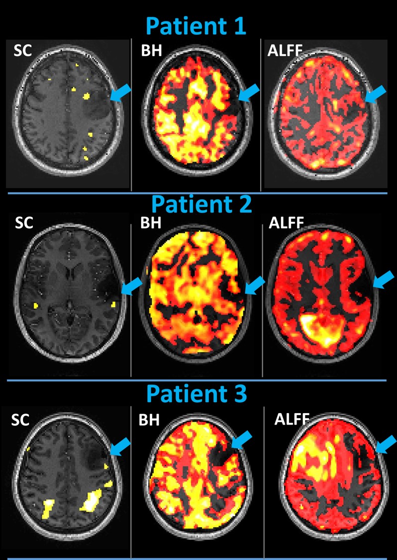
Figure 1:
Three maps (SC language task fMRI activation, BH CVR and rsfMRI ALFF at 7T) overlaid
on T1 structural images are displayed. In Patient
1, expected dorsolateral prefrontal cortex (DLPFC) activation along the
lateral tumor margin is absent. In Patient
2, expected Wernicke’s area (superior temporal gyral) receptive language
activation is reduced along the lateral tumor margin. In Patient 3, expected DLPFC activation is reduced along the lateral
tumor margin. Regionally decreased BH CVR and ALFF in these regions of markedly
reduced activation in these right handed, left language dominant patients is suggestive
of neurovascular uncoupling.