4616
High-b Fast Advanced Spin Echo Diffusion-Weighted Imaging in the Abdomen1Advanced Biomedical Imaging Research Center, Kobe University Graduate School of Medicine, Kobe, Japan, 2Center of Radiology and Radiation Oncology, Kobe University Hospital, Kobe, Japan, 3Toshiba Medical Systems Co., Otawara, Kazakhstan, 4Radiology, Kobe University Graduate School of Medicine, Kobe, Japan
Synopsis
High-b FASE-DWI can improve image quality and decrease image distortion without hampering abdominal lesion detection and ADC measurement.
INTRODUCTION & PURPOSE
In current abdominal MRI, DWI is one of the most important techniques and routinely used worldwide. However, problems still remain such as poor image quality and distortion due to air in lung or intestinal, or outside the body, especially when using single-shot EPI at 3T. To solve these problems, we developed Fast Advanced Spin Echo (FASE)-DWI (WIP). Recently, FASE-DWI at high-b value such as 1000 became available.
The purpose of this study was to assess FASE-DWI at high-b value in evaluation of abdominal diseases.
METHODS & MATERIALS
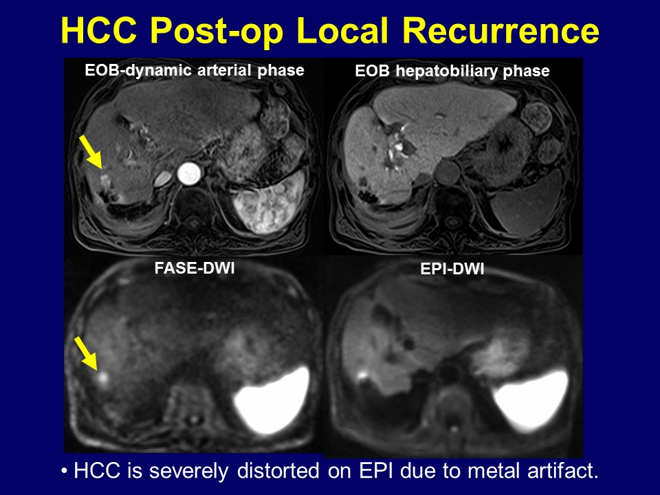
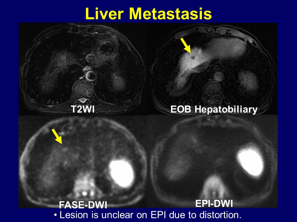
Eighty patients (54 men and 26 women, mean: 67.4 years), who were suspected to have hepato-biliary-pancreatic malignancy and underwent 3T-MRI, were enrolled. 49 malignant lesions (HCC: 27, Liver meta: 10, Bile duct Ca: 3, Panc Ca: 2, pNET: 2, Colon Ca: 2, LN meta: 2, IPMC: 1) were confirmed.
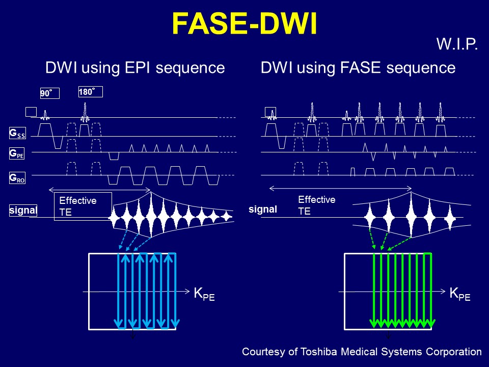
FSE-T2WI, SE-EPI-DWI (TR/TE/FA=6500-7000/70/90, b values: 0 & 1000, matrix: 128 ×128, thickness: 8mm, NEX: 4 scan time: 5:15min, PASTA + SPAIR, PI: 2.5, MPG: (y, z)), and FASE-DWI (TR /TE/FA=10000/74/90, b values: 0 & 1000, matrix: 96 ×112, thickness: 8mm, NEX: 6 scan time: 6:39min, SPAIR, PI: 2.5, MPG: (x, y, z)) were obtained in all patients.
Amount of abdominal gas and ascites on images were recorded for each patient using a 5-point scale. Anteroposterior (AP) and right-to-left (RL) abdominal diameters were measured for each sequence and each patient on the slice with most severe image distortion, and correlation analyses were performed among the sequences. Overall image quality and severity of image distortion were visually assessed using a 5-point scale on EPI-DWI and FASE-DWI, and compared. Regression analyses were done to estimate factors for low image quality and severe distortion. Malignant lesion detection for each patient and conspicuity for each lesion was separately assessed on EPI-DWI and FASE-DWI by two readers using a 5-point scale. Consensus was made and detection was compared using ROC analysis. Apparent diffusion coefficients (ADC) in malignant lesions and background organs were measured on FASE-DWI and compared.
RESULTS
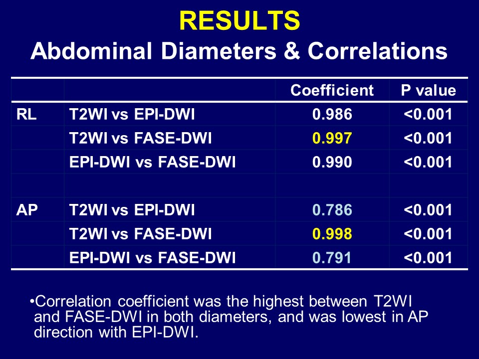
Correlation coefficient was the highest between T2WI and FASE-DWI in both diameters, and was lower in AP direction with EPI-DWI, indicating less image distortion on FASE-DWI. There was no significant difference between overall image qualities (EPI:3.1 vs FASE:3.3, p=0.070). Image distortion was significantly severer on EPI-DWI (3.9 vs 1.3, <0.0001). Sex and gas were found to be significant factors for quality on EPI-DWI (p=0.026, 0.001), and sex, gas, RL diameter were for distortion (0.002, 0.0003, 0.002). Sex was a significant factor for image quality on FASE-DWI (0.012). There was no significant difference in malignant lesion detection (Az: 0.950 vs 0.934) and conspicuity (4.55 vs 4.51). Malignant lesion ADC was significantly lower than background ADC (lesion:1.50 vs background:2.84 x10⁻⁶, <0.0001).SUMMARY
- Distortion was reduced on FASE-DWI.
- No significant deference was found in image quality between the techniques.
- Image quality and distortion on EPI-DWI were significantly affected by abdominal gas.
- Diagnostic performance and conspicuity for malignant lesions were similar on both images.
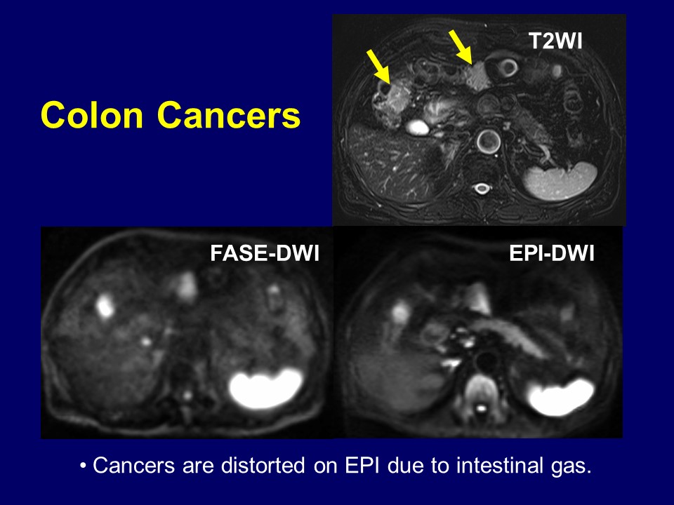
- FASE-DWI was useful in the abdominal parts near air.
CONCLUSION
High-b FASE-DWI can improve image quality and decrease image distortion without hampering abdominal lesion detection and ADC measurement.Acknowledgements
No acknowledgement found.References
Previous reports on extracrainal FASE-DWI
- Ohno Y, et al. EJR 2015 (Lung CA, N-staging)
- Kito S, et al. Oral Surg Oral Med Oral Pathol Oral Radiol Endod 2006(head & neck abscesses)
Figures