4609
Diagnostic Accuracy of Liver Imaging Reporting and Data System 2017 (LIRADS) Criteria for Hepatocellular Carcinoma1Radiology, Well Cornell Medical Center, New york, NY, United States, 2Columbia university medical center, New york, NY, United States
Synopsis
The aim of our investigation is to assess the diagnostic accuracy of the LI-RADS 2017 criteria at two institutions using multiple readers with varying levels of experience with explant or imaging follow-up as the reference standard. Radiology databases from two academic institutions were searched (2013-2014) for patients with a clinical diagnosis of chronic liver disease and at least one reported hepatic observation on dynamic contrast enhanced CT or MRI. This yielded a final cohort of 103 patients with 141 hepatic observations. Two radiologists reviewed the imaging independently and assigned a LI-RADs category to each observation. Inter-reader reliability for LI-RAD assessment was moderate (ICC = 0.63). Sensitivity of LI-RADs categorization for diagnosing HCC was 62% and 59% and specificity was 96% and 84% for reader 1 and 2 respectively. LI-RADs categorization using gadoxetate disodium MR demonstrated higher specificity for HCC diagnosis for reader 2, than MR with extracellular agent.
Introduction
The Liver Imaging Reporting and Data System (LI-RADs) criteria, provides a standardized lexicon and categorizes lesions on an ordinal scale to relay the likelihood of hepatocellular carcinoma (HCC) on imaging (1,2). Initially launched in 2011, the most recent update was performed in 2017, and includes changes to LR categories and criteria (1,2). However, despite being adopted by many, few studies have performed validation of LI-RADS criteria, with current studies limited by single-center cohorts without standardized MR parameters (3-5). Therefore, the aim of our investigation is to assess the diagnostic accuracy of LI-RADS 2017 criteria at two institutions using multiple readers with varying levels of experience with explant or imaging follow-up as the reference standard.Methods
In this IRB approved retrospective study with waiver of consent, radiology databases from two academic institutions were searched for patients with a clinical diagnosis of chronic liver disease and at least one reported hepatic observation on dynamic contrast enhanced CT or MRI performed between 2013-14. CT and MRI images were performed on different scanners with protocols and technical parameters in accordance with LI-RADs 2017 specifications. MRI was performed with either weight-based extracellular contrast (n = 76) or fixed dose hepatobiliary contrast (n = 49). Explant histopathology or biopsy results were reviewed and correlated with imaging findings by the study coordinator, a board certified abdominal radiologist not involved in study interpretations in consultation with a hepatobiliary pathologist (3 yrs experience). Lesions that were completely necrotic and without residual findings to confirm HCC at pathology were excluded. In patients without pathology (n=45), MRI or CT follow-up for at least two years was used as the reference standard. This yielded a final cohort of 103 patients with 141 hepatic observations. Two board certified, fellowship trained abdominal radiologists (R1>15 yrs, R2>2 yrs experience) reviewed the imaging independently in a blinded fashion on a PACS workstation, assigned a LR category, and evaluated major and ancillary features using a standardized worksheet with location maps provided for lesion identification. Statistical analysis included Intraclass correlation for readers, sensitivity, specificity, NPV, PPV and diagnostic accuracy of LI-RADS category per reader.Results
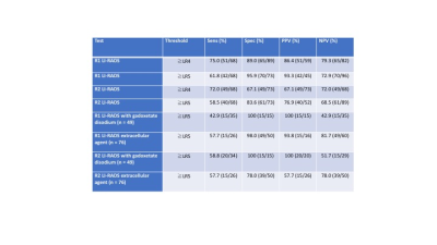
16 observations were evaluated with CT and 125 with MRI. The final diagnoses were: 68 HCCs, 2 intrahepatic cholangiocarcinomas, 2 epitheliod hemangioendotheliomas, 1 fibrolamellar HCC, and 68 benign lesions (23 on pathology and 45 on follow-up imaging). Considering all categories, agreement for LI-RADS category assignment was good with ICC of 0.63 (95% CI: 0.49, 0.71). Based on the expert reader’s (R1) interpretations, none of the 16 LI-RADS category 1 or 2 observations, 8/53 (15%) of 53 category 3 observations, 15/21 (71%) category 4 observations, 36/38 (95%) category 5 lesions, 6/7 (86%) category LR-TIV observations, and 3/6 (50%) category LR-M observations were diagnosed as HCC. Table 1 shows the diagnostic performance of the two readers. Sensitivity was increased and specificity decreased for both readers when using LR 4 and 5 together as definitive for HCC compared to LR5 only. LI-RADs categorization using gadoxetate disodium MR demonstrated higher specificity for HCC diagnosis for R2, than MR with extracellular agent.Conclusions
The observed inter-reader reliability is slightly lower than what has been observed in previous studies (6), however, our study cohort represents consecutive patients presented in a fashion that would be encountered in real world clinical practice with radiologists of different experience levels. The observed diagnostic accuracy is comparable to a prior study which evaluated ≤ 20 mm lesions detected on antecedent ultrasound, however, the higher sensitivity in our study is likely due to inclusion of all observations regardless of size (4). LI-RADS categorization with gadoxetate disodium for R2 demonstrated improved specificity and similar sensitivity than with extracellular agents and therefore, gadoxetate disodium may have the potential to provide increased diagnostic accuracy for more inexperienced readers.Acknowledgements
No acknowledgement found.References
References: [1] Mitchell DG et al. Hepatology 2015;61(3):1056-65. [2] Elsayes KM et al. J Hepatocell Carcinoma 2017;4:29-39 [3] Ehman EC et al. Abdominal Radiology 2016;41(5):963-9 [4] Darnell A et al. Radiology 2015;275(3):698-707 [5] Choi SH et al. Investigative radiology. 2016;51(8):483-90. [6] Fowler KJ, et al. Radiology 2017;[Epub ahead of print].Figures

