4597
Preliminary study of evaluating Intrarenal Oxygenation in Iodinated Contrast-Induced Acute Kidney- Injured pig models by BOLD MRI1TianJin 4th Center Hospital, TianJin, China, 2Philips Healthcare, Beijing, China
Synopsis
Iodinated contrast
agent (CA) is important for many radiologic and interventional procedures.
However, contrast-induced acute kidney injury (CIAKI) is potentially
life-threatening, especially for patients with impaired renal function, and it has
become one of the leading causes of hospital-acquired acute kidney injury (AKI)[1] . The pathogenesis of CIAKI is not yet fully understood, but the
relevance of renal medullary hypoxia in the pathophysiology of AKI is well
accepted. In recent years, many studies focused on the change of intrarenal oxygenation
and showed renal hypoxia especially in medulla. Blood oxygen level dependent
magnetic resonance imaging (BOLD MRI) is a noninvasive method that can assess
hypoxia by utilizing the endogenous contrast generated by paramagnetic
deoxyhemoglobin[2] .
Purpose
To evaluate the renal oxygenation determined by blood oxygenation level dependent magnetic resonance imaging (BOLD MRI) in pig models of contrast-induced acute kidney injury (CIAKI).Methods
This study was approved by the institutional animal care and use committee. All MRI examinations were performed under general anesthesia. All pigs were forced to refrain from food and water for 12 hours. Seven pigs (Bama suckling pig) weighting between 80 and 90 kg were injected with ioversol 320 at a flow rate of 3ml/s and a dose of 6ml/kg. Kidney BOLD MRI studies were performed on a 3.0T scanner (Achieva TX, Philips Healthcare, Best, the Netherlands) before the injection of iodine contrast agent and 25 min after the injection, respectively. Kidney BOLD MRI was carried out by using multiple T2*weighted gradient echo sequence. Five slices with 5mm thickness with a gap of 1mm were obtained in the coronal plane as well as following parameters: TR=200ms. TE=2.5-34ms, echo time spacing=4.5 ms. flip angle=45°. Bandwidth=318.6Hz/pixel. Matrix=108×162. FOV=200×304mm. The medullary R2* (MR2*) and cortical R2*(CR2*) values were extracted and quantified on BOLD MRI. Then all the extracted parameters were compared between before the injection of iodine contrast agent and 25 min after the injection using Student’s t test in IBM SPSS Statistics 22.0 (Armonk, New York, USA) where P<0.05 indicated significant different. After MRI study, all pigs were executed and then the pathological changes were observed.
Results
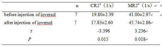
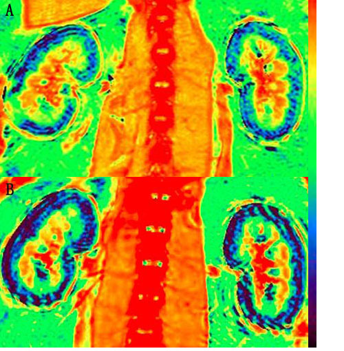
As shown in Table 1 and Figure 1, compared with baseline, the MR2* values were significantly increased 25 min after the injection of ioversol, while the CR2* values were significantly decreased. In both conditions of baseline and 25 min after injection of ioversol, the CR2* values were significantly lower than MR2* values, largely due to the higher concentrations of deoxyhemoglobin in the medulla.
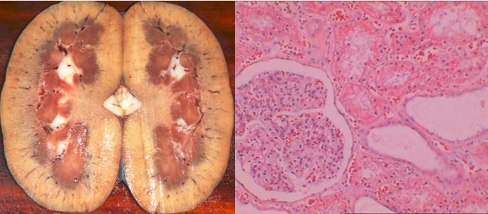
As shown in Figure 2, 25 min after injection of ioversol, we can see the renal tubular cells turbid swelling, vacuole formation, brush edge disintegrating off, and in renal tubular cavity we can find shedding cell debris, renal interstitial edema, vasodilation and congestion. No obvious abnormalities were observed in the Glomerular.
Discussion
Our results showed the R2* value was considerably lower in cortex than that in medulla, which was consistent with previous studies. Compared with baseline, the MR2* values were significantly increased 25 min after the injection of ioversol, it was presumed that the medullary hypoxia was predominantly caused by reduced renal perfusion and enhanced oxygen consumption for tubular reabsorption[3] , which may indicate the early clinical stage of CIAKI. After injection of iodinated contrast agent, the renal tubular damaged, such as cells turbid swelling, vacuole formation, brush edge disintegrating off, and so on. Which is consisted with the pathological changes in CIAKI. Iodinated contrast agent induced alteration of renal hemodynamics and direct tubular damage play a critical role in CIAKI. This is a preliminary study with limited time for recruiting pigs, but we will include more pigs in future for further validation.Conclusion
From our preliminary results, we have found that BOLD MRI can detect and assess the renal hypoxia in CIAKI in the early stage. Hypoxia in medulla was more sensitive than in cortex. And the MR2* values may indicate the renal medulla pathologic damage.Acknowledgements
No acknowledgement found.References
[1] Seeliger E, Sendeski M, Rihal CS, et al. Contrast-induced kidney injury: mechanisms, risk factors, and prevention. Eur Heart J. 2012; 33:2007 -2015.
[2] Zhou HY, Chen TW, Zhang XM. Functional Magnetic Resonance Imaging in Acute Kidney Injury: Present Status. Biomed Res Int. 2016; 2016: 2027370.
[3] Samuel N H, Seymour R, Christian R. Renal parenchymal hypoxia, hypoxia adaptation, and the pathogenesis of radiocontrast nephropathy. Clin J Am Soc Nephrol,2008,3:288-296.
Figures