4595
Tri- and Biexponential Diffusion Analyses of the Kidney: Effect of Respiratory Controlled Acquisition on Measurement Accuracy and Repeatability of Diffusion Parameters1Department of Radiological Technology, Kanazawa University Hospital, Kanazawa, Japan, 2Faculty of Health Sciences, Institute of Medical, Pharmaceutical and Health Sciences, Kanazawa University, Kanazawa, Japan, 3Department of Radiology, Kanazawa University Hospital, Kanazawa, Japan
Synopsis
To investigate the effect of respiratory-controlled acquisition on intravoxel incoherent motion analysis in the kidney, we compared the fitting accuracy and repeatability of diffusion parameters with tri- and biexponential models among three different methods, ie., respiratory triggering, abdominal belt, and free breathing. Respiratory triggering shows better fitting accuracy and repeatability of tri- and biexponential diffusion parameters compared with free breathing. Moreover, abdominal belt can improve the measurement repeatability even with free breathing.
INTRODUCTION
Intravoxel incoherent motion (IVIM) analysis with biexponential function provides both perfusion and diffusion information and is useful to assess renal function and classify renal tumor.1,2 Previous studies have reported that IVIM analysis with triexponential function (assuming three diffusion components: perfusion-related diffusion, fast-free diffusion, and slow-restricted diffusion) provides more detailed information on perfusion and diffusion in tissues.3,4 Moreover, it has been suggested that diffusion signal in the kidney is better fitted with triexponential model than biexponential model.5 However, the clinical application of these analyses in the kidney is hampered because of the low measurement accuracy and repeatability. One possible reason for this limitation is physiological motion mainly caused by respiration resulting in image artifacts, blurring, and signal fluctuation. Therefore, to investigate the effect of respiratory-controlled acquisition on IVIM analysis in the kidney, we compared diffusion parameters with tri- and biexponential models among three different methods, ie., respiratory triggering (RT), abdominal belt (AB), and free breathing (FB).METHODS
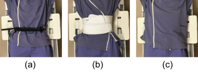
On a 3.0-T MRI, coronal diffusion-weighted images of the kidney were obtained in seven healthy volunteers (all men; mean age, 25.1±2.6 years) using single-shot diffusion echo-planar imaging with RT, AB, and FB (Fig. 1). In AB method, the belt was wrapped tightly around the subject’s abdomen to reduce the respiration-induced motion. The imaging parameters were: repetition time, one respiration cycle (4000-5454 ms) for RT and 4000 ms for AB and FB; echo time, 62.1 ms; acquisition matrix, 96 × 120; b-values, 0 to 1200 s/mm2 (11points); field of view, 360 mm; slice thickness, 5 mm; number of signals averaged, 2 for RT and 3 for AB and FB; and parallel imaging factor, 2. The same scan was repeated twice to assess the repeatability of the measurements. We determined mean signal intensity in the renal cortex at each b-value. Then, perfusion-related, fast-free, and slow-restricted diffusion coefficients (Dp, Df, and Dr, respectively) and fractions (Fp, Ff, and Fr, respectively) were calculated form triexponential fitting. Note that we assigned Df to the literature value of the diffusion coefficient of free water at 37℃ (3.0 × 10⁻³ mm2/s) in the same manner as a previous study.4 Moreover, perfusion-related diffusion coefficient (D*), the fraction (F), and perfusion-independent diffusion coefficient (D) were calculated from biexponential fitting. Root mean square errors for tri- and biexponential models (RMSEtri and RMSEbi, respectively) were obtained to assess the deviation of the fitted to measured data, ie., the fitting accuracy. Repeatability coefficients (RC) were calculated from Bland-Altman plots to assess the repeatability of diffusion parameters. These values were compared among the three methods.RESULTS AND DISCUSSION
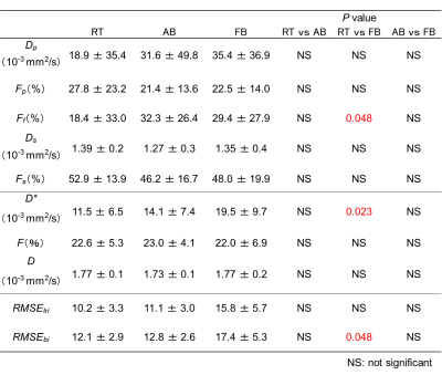
Diffusion parameters and RMSE for each method are presented in Table 1. D*, Ff, and RMSEbi with FB were significantly higher than those with RT, whereas there were no significant differences in the other parameters among the methods. These results indicate that some diffusion parameters with FB are more susceptible to signal variations due to respiration-induced image blurring. Another finding of this study is that the slower diffusion coefficients (Ds and D) calculated at higher b-value range (at least > 200 s/mm2) are robust independent of the use of respiratory-controlled acquisition. Table 2 shows RC of diffusion parameters in the three methods. RT showed the best repeatability for Dp, Ds, Fs, D*, and D, and the RC for Fp, Ff, and F were smallest in AB. FB had the worst repeatability for all diffusion parameters.CONCLUSION
Respiratory triggering shows better fitting accuracy and repeatability of tri- and biexponential diffusion parameters compared with free breathing. Moreover, abdominal belt can improve the measurement repeatability even with free breathing.Acknowledgements
No acknowledgement found.References
1. Gaing B, et al. Invest Radiol. 2015; 50: 144-152.
2. Bane O, et al. J Magn Reson Imaging. 2016; 44: 317-326.
3. Hayashi T, et al. J Magn Reson Imaging. 2013; 38: 148-153.
4. Ohno N, et al. J Magn Reson Imaging. 2016; 43: 818-823.
5. Baalen S, et al. J Magn Reson Imaging. 2017; 46: 228-239.
Figures


Table 2. Repeatability coefficient of tri- and biexponential diffusion parameters in each method