4589
Optimizing Diffusion-Weighted MRI of the Kidneys: Comparison between Simultaneous Multi-Slice and Integrated Slice-by-Slice Shimming Echo Planar Sequences1Radiology, Peking Union Medical College Hospital, Beijing, China, 2MR Collaboration, Siemens Healthcare Ltd., Beijing, China., Beijing, China, 3Siemens Healthcare, Application Development, Germany, Erlangen, Germany, 4Nephrology, Peking Union Medical College Hospital, Beijing, China
Synopsis
This study aimed to evaluate the image quality and scan time of the simultaneous multi-slice (SMS) technique compared to integrated slice-by-slice shimming (iShim[MB1] ) single-shot echo planar imaging (ss-epi) for diffusion-weighted imaging (DWI) of the kidneys. Six healthy subjects and 22 patients with renal disease underwent both SMS and iShim DWI scans on a 3T MR scanner. Despite the average scan time of SMS DWI being decreased by 53.5% compared to iShim DWI, the image quality of SMS DWI was slightly better, with a higher signal-to-noise ratio and similar contrast-to-noise ratio. ADC values of the kidneys were comparable in both DWI sequences. [MB1]Our product name is "SliceAdjust".
Introduction
Kidney diseases are very common. Diffusion-weighted imaging (DWI) is an important magnetic resonance imaging (MRI) technique to evaluate kidney disease. Despite its potential applications, however, DWI has not been part of most routine clinical kidney protocols. One of the primary reasons is the relatively long acquisition time. With the advent of the simultaneous multi-slice (SMS) technique, scan time reduction for kidney DWI may become feasible, but it remains unknown whether the image quality of SMS DWI would be compromised compared with other advanced DWI imaging techniques such as integrated slice-by-slice shimming (iShim) single-shot echo planar imaging (ss-epi) sequences. The purpose of this study was to compare the performance of SMS and iShim of kidney DWI for scan time and image quality.Method
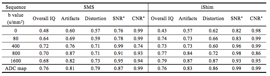
Data were collected on a MAGNETOM Skyra 3T MR scanner (Siemens Healthcare, Erlangen, Germany) using an 18-channel phased-array body coil combined with a 32-channel spine coil. Six healthy volunteers (3 males, 3 females; mean age, 29 ± 6 years) and 22 patients (14 males, 8 females; mean age, 42 ± 14 years) with confirmed diagnoses of kidney diseases (Gitelman syndrome, n = 4; IgA nephropathy, n = 7; malignant hypertension, n = 2; chronic kidney disease, n = 9) were included in the study. Axial DWI of the kidneys was performed using the prototype SMS sequence followed by the prototypic ss-epi sequence with iShim, with the following parameters: TR/TE = 3000/66 ms (SMS), 6400/64 ms (iShim), Fat suppression mode: SPAIR (SMS), Water excitation (Ishim), FOV = 400 × 325 mm2, slice thickness = 4 mm, 30 slices, voxel size = 1.6 × 1.6 × 4.0 mm3, GRAPPA (PE) 2, and b-values = 0, 80, 400, 800, 1600 s/mm2. Scan times: 1 min 38 s for SMS DWI, and 3 min 33 s for iShim DWI; and the slice acceleration factor was 2 for SMS. The subjective image quality of five b-values and ADC maps was evaluated by two experienced radiologists independently. A 5-point scale was used to assess the overall image quality, occurrence of artifacts, and distortion, with “5” indicating excellent or no artifacts or distortion. The signal-to-noise ratio (SNR), contrast-to-noise ratio (CNR), and ADC values were calculated by using the five b values of the kidney, measured for each DWI sequence. All the measurements were performed twice by two experienced radiologists. Inter-observer agreement for image quality assessment was analyzed.Results
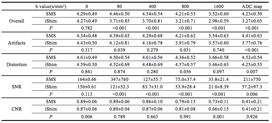
The inter-observer agreement for the SNR and CNR was good to excellent, whereas the inter-observer agreement for the subjective image quality scores was slightly compromised on the b0 images but good to excellent on the other four b values and ADC maps (Table 1). On the b0 images, no statistically significant differences in SNR or the subjective image quality scores between SMS and iShim DWI were observed, but the CNR was significantly higher in SMS compared to iShim DWI. On the other four b-value images, the SNR and overall image quality scores were higher in SMS DWI, but the score for image distortion was comparable between SMS and iShim DWI. On b80 and b800 images, SMS DWI had fewer artifacts, but this difference was not observed on b400 and b1600 images. The CNR was significantly higher in SMS DWI on the b1600 images (0.73 ± 0.11 vs. 0.66 ± 0.15, P=0.001) but was similar between SMS and iShim DWI on b80, b400, and b800 images (P>0.05). Regarding the ADC maps, the SNR and all the subjective image quality scores were significantly higher in SMS DWI, whereas the CNR showed no significant differences between SMS and iShim DWI. The ADC values of the kidneys were similar in SMS and iShim DWI (1.54 ± 0.11 × 10⁻³ mm2/s vs. 1.52 ± 0.16 ×10⁻³ mm2/s, P=0.343). Details of the image quality assessment are summarized in Table 2.Discussion and Conclusion
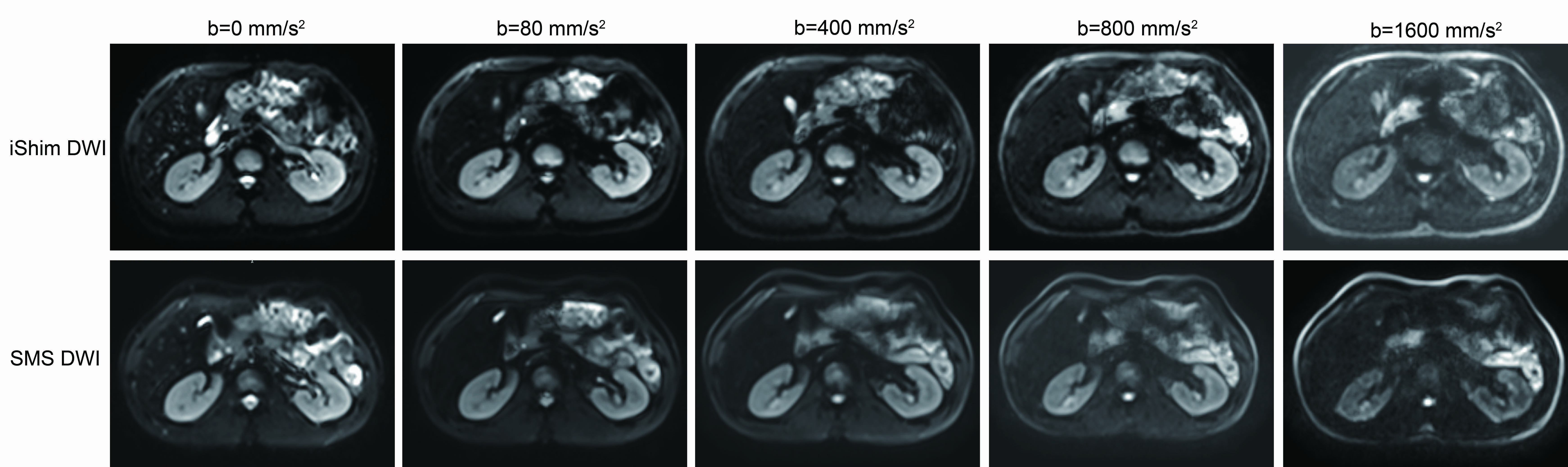
This is the first study comparing the image quality of two advanced DWI imaging techniques, SMS and iShim DWI, for evaluating the kidneys systematically. Compared with the iShim DWI sequence, SMS DWI of the kidneys can substantially reduce scan time without compromising SNR, CNR, or image quality while maintaining the stability of the ADC quantification (Figure 1). Previous studies1,2 have reported that iShim can improve the quality in the imaging region that has strong susceptibility artifacts. In our study, the results showed that this effect was not so strong in the kidney. With a shorter scan time, SMS DWI may contribute to a more efficient workflow in clinical practice and might also be applied in diffusion schemes with long acquisition time, such as intra-voxel incoherent motion. Further studies are needed to investigate the application of SMS DWI for evaluating different renal pathologies.Acknowledgements
NoReferences
1. Zhang H, Xue H, Alto S, et al. Integrated Shimming Improves Lesion Detection in Whole-Body Diffusion-Weighted Examinations of Patients With Plasma Disorder at 3 T. Invest Radiol.2016; 51:297-305.
2. Sven S. Walter, Wei Liu, et al. Combination of integrated dynamic shimming and readout- segmented echo planar imaging for diffusion weighted MRI of the head and neck region at 3Tesla . Magnetic Resonance Imaging. 2017;42:32-36.
Figures