4575
High dielectric material application in fetal MR imaging at 3T1Lauterbur Research Center for Biomedical Imaging, Shenzhen Institutes of Advanced Technology, Chinese Academy of Sciences, Shenzhen, China, 2Shenzhen Key Laboratory for MRI, Shenzhen, China, 3School of Basic Science, Guangzhou Medical University, Guangzhou, China, 4Department of Radiology and Biomedical Imaging, University of California San Francisco, San Francisco, CA, United States, 5UCSF/UC Berkeley Joint Graduate Group in Bioengineering, San Francisco, CA, United States
Synopsis
In this paper, we evaluated the imaging performance in fetal imaging for supine and lateral positions of pregnant patients with/without high permittivity dielectric pad at 3T through numerical modeling and simulation. The results suggest that the lateral position is advantageous over the supine positions in terms of B1+ efficiency and SAR or MR safety.
Introduction
Fetal magnetic resonance imaging (MRI) has become a hot spot in recent years due to its advantages over other imaging modalities such as large field of view, high spatial resolution and high soft-tissue contrast1. It has been shown that high dielectric material has the ability to change the electromagnetic field distribution, and increase the imaging sensitivity and safety2,3. These features could be significantly beneficial for fetal MRI. Because of the high density and heavy weight, the high dielectric material is often placed on the patient table. During the examination, pregnant patients usually take supine or lateral positions. However, effects of the dielectric materials to imaging performance for these two possible positions have not been quantitatively evaluated. In this work, we modeled and numerically evaluated two typical positions of pregnant patient in MR scans and investigated the effect of high dielectric material to fetal imaging at 3T. The parameters evaluated include position of the high dielectric material, the distribution of the B1+ field, and SAR.Methods and Materials
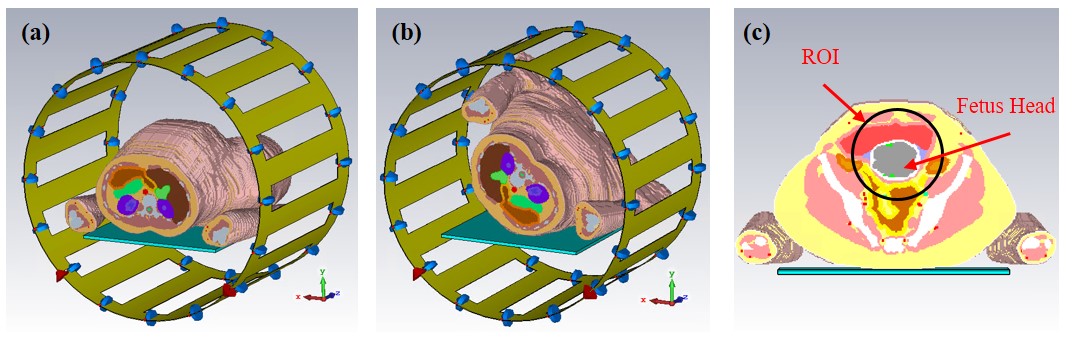
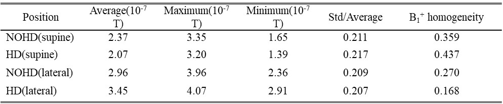
We investigated the influences of different human scan position by the numerical methods for calculating electromagnetic field. The commercial CST software (Darmstadt, Germany) is used to simulate the models. A high dielectric pad was designed to represent a very dense mixture of 4:1 barium titanate-to-water (mass/ mass ratio)4. In our design, the high dielectric pad with dimensions of 400mm×350mm×7.5mm was placed on the patient table. The relative permittivity and the electric conductivity of the pad were respectively set to 220 and 0.473 S/m 4. A quadrature 16-element high-pass birdcage coil with 312.5mm-radius end-ring and 470mm in length tuned to 123.2MHz using capacitor of 66pF in each gap was modeled as a volume coil. The model of human tissue imported from pregnant woman with 387mm length in CST was placed in the center of the birdcage coil with supine position as shown in Fig 1(a). In the modeling, the lateral position was obtained by rotating the supine human tissue with 140 degree along the z-axis. The two discrete excitation ports orthogonally distributed on the side of end ring as shown in Fig 1. The number of mesh cells was about 7.5 million, and the boundary conditions were set as open in all direction. The models were computed using two GPU accelerator card with/without the high dielectric pad load. Time-domain solver was employed, and the steady-state accuracy limit was set to -30 dB. All the post process results were normalized to the input power of 1W and calculated by using MATLAB (Mathworks, Natik, MA, USA) code. In order to consider the value of the B1+ field in the area of fetus head, the transverse plane in the center of volume coil was selected and the black circle radius marked as ROI was 60mm as shown in Fig. 1(c). The maps of SAR were calculated using 10g average mass and IEEE/IEC62704-1 as average method. The maximum value of SAR corresponds to all the human tissue. The homogeneity of B1+ field in the ROI was calculated as:
B1+ homogeneity = ( B1+Max - B1+Min )/( 2×B1+Average )
The transmit efficiency in the ROI was calculated as:
Transmit Efficiency = B1+Average/Total SAR
Results
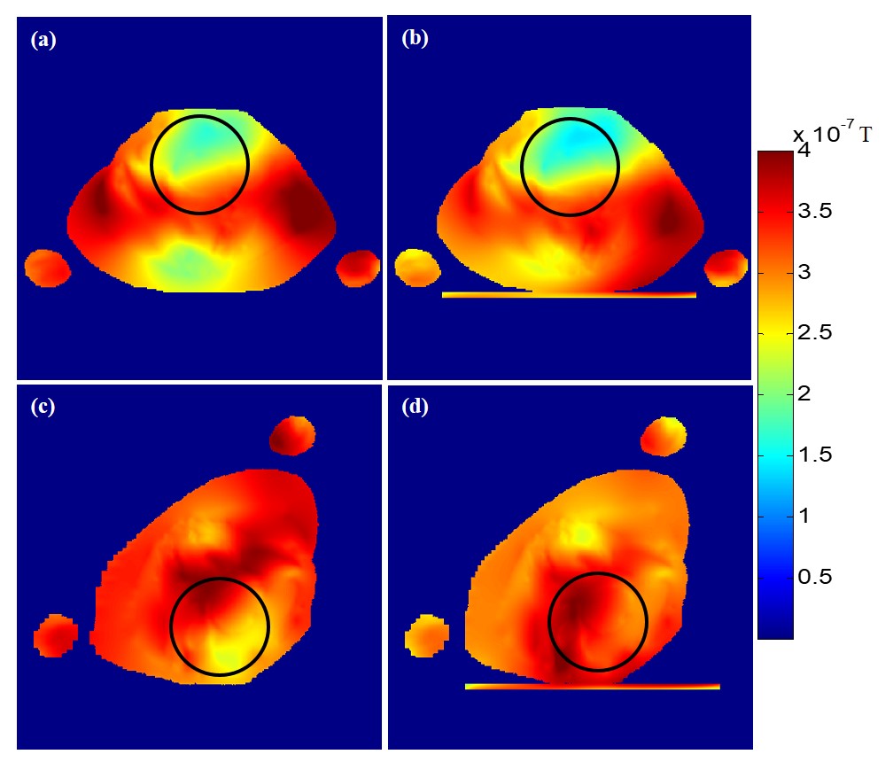
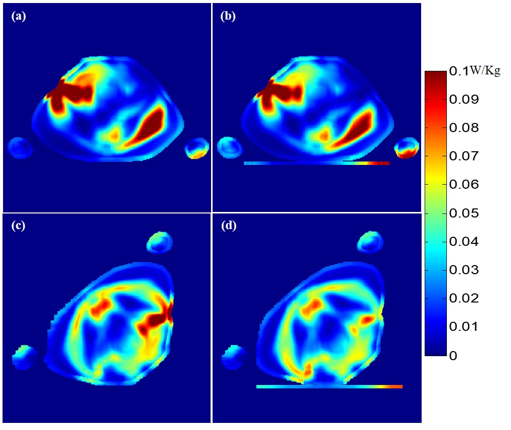
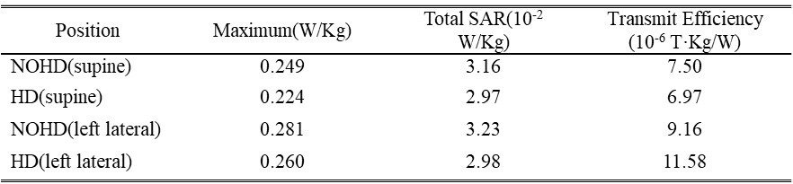
As shown in Fig. 2 and Fig 3, the B1+ and SAR field maps were calculated in transverse plane corresponding without/with the high dielectric pad load in supine and lateral position. The average and the maximum value of the B1+ field in ROI of human tissue were increased when using the lateral position. Measurements of the B1+ average value and maximum value in ROI showed a significant improvement when using lateral position. The average value of B1+ field, the A, the B and the total SAR with lateral position increase respectively by 16.6%, -37.8%, -34.4%, and -7.7% when using the pad. As shown in Table I and Table II, the lateral position is much more effective for creating a local increase of the transmit efficiency and for improving the B1+ homogeneity in ROI when compared to supine position. Additionally, the total SAR in human tissue was at nearly the same level for two positions.Discussion/Conclusion
From the results in this study, high permittivity dielectric material is able to change the distribution of B1+ field in patients for both positions. Shorter distance between high permittivity dielectric material and the imaging target could help to increase the B1+ field in ROI. This study, therefore, suggests that the lateral position would be advantageous over the supine position in improving the B1+ field efficiency and reducing SAR in ROI in the fetal MR imaging.Acknowledgements
This work is supported in part by National Natural Science Foundation of China No. 81527901, 81327801, national key R&D program no. 2016YFC0100100, national grants no. 51307171, 61571433, 61401450, 81470077, 81571669 and 2013CB733800/2013CB733803, provincial grants no. 2014B030301013, 2015B020214006 and 2014A030310200, city grant no.KQJSCX20160301143250, CYJ20140417113430589, JSGG20141020103440414,KQCX2015033117354154, internal grant no. 201314, and a Pengcheng Scholar Award.References
[1] Wielandner Alice, et al. Potential of magnetic resonance for imaging the fetal heart. Seminars in Fetal and Neonatal Medicine. Vol. 18. No. 5. WB Saunders, 2013. [2] Koolstra, Kirsten, et al. Improved image quality and reduced power deposition in the spine at 3T using extremely high permittivity materials. Magnetic Resonance in Medicine (2017). [3] Luo Minmin, et al. Numerical assessment of the reduction of specific absorption rate by adding high dielectric materials for fetus MRI at 3T. Biomedical Engineering/Biomedizinische Technik 61.4 (2016): 455-461. [4] Brink, Wyger M. et al. High permittivity pads reduce specific absorption rate, improve B1 homogeneity, and increase contrast‐to‐noise ratio for functional cardiac MRI at 3T. Magnetic resonance in medicine 71.4 (2014): 1632-1640.Figures