4549
Calculation of the Susceptibility Effect on the Rate of Transverse Relaxation using Real Microvascular Networks1Department of Biomedical Engineering, Boston University, Boston, MA, United States, 2Department of Radiology, Athinoula A. Martinos Center for Biomedical Imaging, Charlestown, MA, United States, 3Division of Health Sciences and Technology, Massachusetts Institute of Technology, Cambridge, MA, United States, 4Departments of Radiology, UC San Diego, La Jolla, CA, United States, 5Departments of Neurosciences, UC San Diego, La Jolla, MA, United States
Synopsis
We obtain the exponent in the power law relation of the transverse relaxation rate and the susceptibility difference between vessels and tissue from first-principles calculations using our recently developed VAN model. We find that this exponent is close to 1, and is more uniformly distributed across
Introduction
The relation between the effective transverse relaxation rate $$$R2^*$$$ and the susceptibility difference $$$\Delta \chi$$$ between blood and tissue can be written as $$$\Delta R2^*\propto CBV\Delta\chi^{\beta}$$$. The main mechanism that introduces a non-linear susceptibility effect ($$$\beta\neq 1$$$) is proton diffusion. Early studies have found that $$$\beta=1$$$ around large vessels and $$$\beta=2$$$ around small vessels [1]. An estimation of the value of $$$\beta=1.5$$$ was obtained from Monte-Carlo simulations of protons diffusing around a random-cylinders approximation of the cortical microvasculature performed over 20 years ago [1, 2]. However, these studies have not taken into account the complex structures of a real vascular network. More importantly, the heterogeneous distribution of deoxy-hemoglobin in different compartments and vessel sizes has not been analyzed. As such, the value of $$$\beta$$$ relevant for BOLD fMRI needs a more detailed investigation. A characterization of the field strength dependence of $$$\beta$$$ is also desired due to more studies being conducted at ultra-high-fields ($$$B_0=7T$$$ and above). Methods
We utilized our vascular anatomical network model to compute MR signals at 5 different magnetic field strengths for 6 vascular stacks and fixed $$$TE=30 ms$$$ to ensure the same diffusion length. We obtain the value of $$$\beta$$$ from fitting the $$$lnR2^*$$$ vs. $$$ln\Delta\chi$$$ curve by varying input $$$\Delta\chi$$$. The uniform vascular partitioning sets $$$\Delta\chi$$$ to be uniformly distributed within vessels and the deoxyhemoglobin-weighted vascular partitioning is using a heterogeneous $$$\Delta\chi(\vec{r})=\Delta\chi Hct(1-SO_2(\vec{r}))$$$, where Hct is the hematocrit and $$$SO_2$$$ is the oxygen saturation. We also use Monte-Carlo simulations using random-cylinders models to understand the vessel size dependence of $$$\beta$$$ at different field strengths.Results
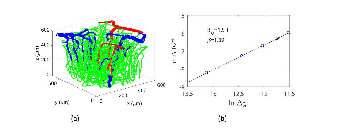
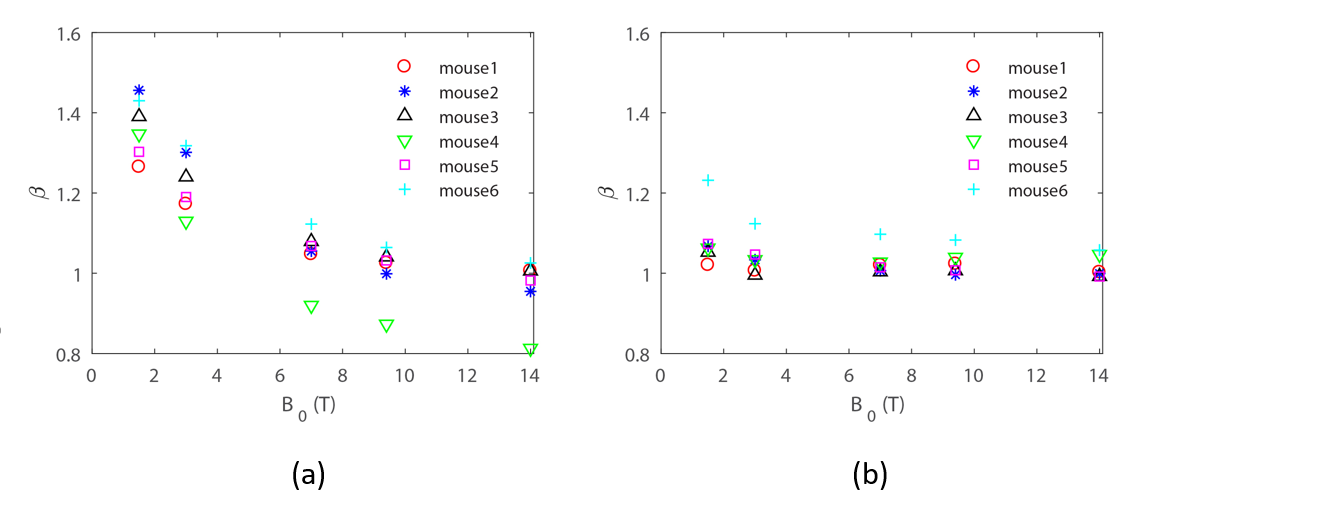
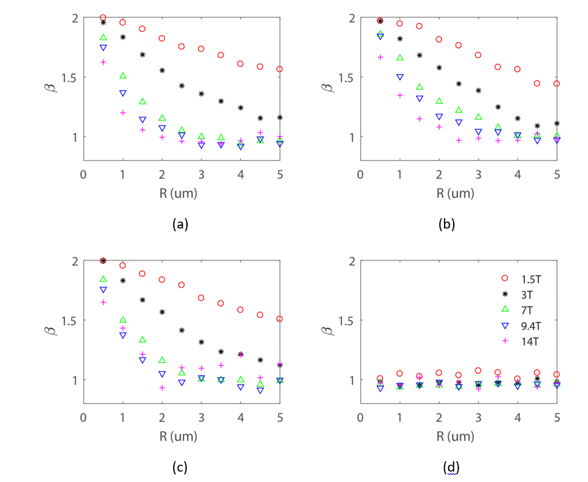
Fig. 1 shows an example of a vascular stack used in our simulations and its corresponding $$$\beta$$$ value obtained from the slope of the $$$lnR2^*$$$ vs. $$$ln\Delta\chi$$$ curve. Fig. 2 a and b show $$$\beta$$$ obtained from uniform vascular partitioning and deoxyhemoglobin-weighted vascular partitioning of $$$\Delta\chi$$$, respectively. $$$\beta$$$ can vary across regions and subjects and decreases with magnetic field strength. At high field strength, $$$\beta$$$ is more uniformly distributed, which is crucial for the accuracy of the CBV measurements using contrast agents that assumes $$$\Delta R2^*\propto CBV$$$. Fig. 3 shows the value of $$$\beta$$$ obtained from Monte-Carlo simulations of random cylinders. We see that at $$$B_0=7 T$$$ and above, the value of $$$\beta$$$ is close to 1 for capillaries $$$r\approx 3\mu m$$$, so that the average $$$\beta$$$ for different vascular stacks is more uniform and can be set to 1. For the BOLD response $$$\beta$$$ can be set to 1 at $$$B_0=3 T$$$ and above, as opposed to $$$\beta=1.5$$$ from earlier studies.Conclusion
We have analyzed the susceptibility effect on the transverse relaxation rate using real micro-vascular networks using our recently developed VAN modeling. Both the uniform vascular partitioning and the deoxyhemoglobin-weighted vascular partitioning of $$$\beta$$$ are studied. We show that the parameter $$$\beta$$$ which governs the dependence of the transverse relaxation rate on the magnetic susceptibility shift is closer to the large vessel limit $$$\beta=1$$$ at higher field strength. Our work provides insights on the fundamental question of the impact of proton diffusion on MR signals at different field strengths as well as practical applications for CBV measurements with contrast agents and rCMRO2 measurements with BOLD fMRI.Acknowledgements
This work was supported in part by the NIH NIBIB (grants P41-EB015896, and R01-EB019437), by NIH R01-MH111359, by the BRAIN Initiative (NIH NIMH grant R01-MH111419), and by the MGH/HST Athinoula A. Martinos Center for Biomedical Imaging.References
1. Boxerman, J.L., Hamberg, L.M., Rosen, B.R., Weisskoff, R.M., 1995. MR contrast due to intravascular magnetic susceptibility perturbations. Magn. Reson. Med. 34, 555–566.
2. Davis, T.L., Kwong, K.K., Weisskoff, R.M., Rosen, B.R., 1998. Calibrated functional MRI: mapping the dynamics of oxidative metabolism. Proc. Natl. Acad. Sci. 95, 1834–1839.
Figures