4547
Test-Retest Reproducibility of Cerebral Blood Flow at Rest and During A Vigilance Task1Center for functional Neuroimaging, University of Pennsylvania, Philadelphia, PA, United States
Synopsis
It is unclear whether ASL test-retest reproducibility would be different between absolute CBF and relative task-induced CBF changes, and across resting and task scans. Here we scanned 15 healthy participants three times in a 5-day well-controlled study while participants were at rest and performing a simple vigilance task. Absolute CBF showed excellent test-retest reliability across three days, which were comparable to performance reliability. However, reliability of task-induced CBF changes were much lower than reliability of absolute CBF, suggesting that absolute CBF rather than relative CBF changes may be a more reliable brain function measurement for longitudinal and clinical studies.
Introduction
Quantitative cerebral blood flow (CBF) measurements from arterial spin labeling (ASL) perfusion imaging is increasingly used to access regional brain function in cognitive and clinical neuroscience studies 1,2. Previous studies have consistently demonstrated high test-retest reliability of CBF over time 3-5. However, it is unclear whether the reproducibility of CBF would be different between absolute CBF and task-induced relative CBF changes, and across resting and task scans. It is also unknown whether the reliability of CBF measures would be comparable to the reliability of behavior performance. In this study, we aimed to quantify test-retest reliability of absolute CBF measurements and relative task-induced CBF changes both at rest and during performing a simple and reliable psychomotor vigilance test (PVT).Methods
We analyzed ASL imaging data from 15 healthy control subjects (6 females, mean age = 35±9 years) from a strictly controlled 5-day and 4-night in-laboratory sleep study. Participants slept 8-9 hours every night in the laboratory. Their sleep and waking activities, diet behavior, and cognitive performance were continuously monitored throughout the study. Each subject were scanned three times on the morning of days 2, 3, and 5, when they were performing a 10-minute PVT and at resting states before (Rest1) and after (Rest2) the PVT. ASL imaging data were acquired using pseudo continuous ASL (pCASL) sequence and analyzed using SPM8 and fMRI Groccer toolbox. Both voxel-wise and region-of-interest (ROI) analyses were conducted. Intra-class correlation coefficients (ICC) were calculated to quantify test-retest reliability across the scans.Results
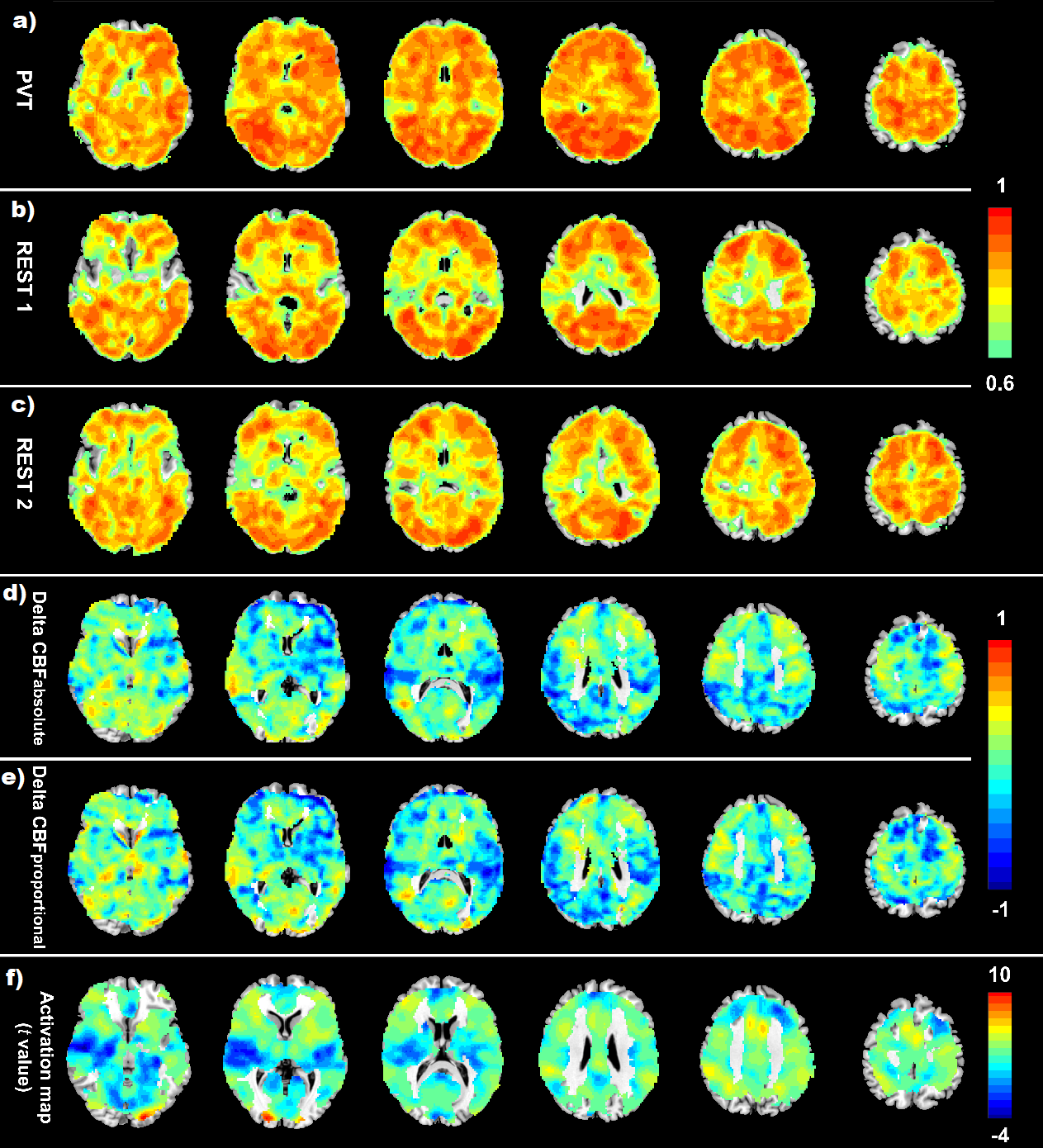
The PVT performance demonstrated excellent test-retest reliability (ICC values ranged from 0.87 to 0.94) across three scans. Voxel-wise analysis of the absolute CBF values also showed excellent test-retest reliability for both resting and PVT scans (Fig.1a-c), with the ICC values slightly higher during the PVT (median ICC = 0.93) than at rest (median ICC = 0.91). However, the test-retest reliability of relative task-induced CBF changes was much lower from both voxel-wise (Fig.1 d-e) and ROI analyses, with ICC values ranging from -0.28 to 0.45 for the PVT-induced activation and deactivation clusters.Conclusions
Consistent with previous studies, absolute CBF showed excellent test-retest reproducibility across three scans both at rest and during the PVT. The ICC values of absolute CBF were comparable to those of PVT performance. However, task-induced regional CBF changes showed much lower test-retest reproducibility, suggesting that reliability of PVT performance may not be fully explained by reliability of task-induced activation or deactivation. These findings suggest a preference of using absolute CBF over relative task-induced CBF changes as a biomarker for assessing regional brain function, especially for longitudinal and clinical studies.Acknowledgements
This research was supported in part by NIH grants R01 HL102119, R01 MH107571, P30 NS045839, and Parkinson's Foundation translational research grants.References
1. Detre JA, Wang J, Wang Z, et al. 2009. Arterial spin-labeled perfusion MRI in basic and clinical neuroscience. Curr Opin Neurol. 2009; 22, 348-355.
2. Detre JA, Rao H, Wang DJ, et al. Applications of arterial spin labeled MRI in the brain. J Magn Reson Imaging. 2012; 35: 1026-1037.
3. Chen Y, Wang DJ, Detre JA. Test-retest reliability of arterial spin labeling with common labeling strategies. J Magn Reson Imaging. 2011. 33, 940-949.
4. Zou Q, Miao X, Liu D, et al. Reliability comparison of spontaneous brain activities between BOLD and CBF contrasts in eyes-open and eyes-closed resting states. NeuroImage. 2015; 121, 91-105.
5. Steketee RM, Mutsaerts HJ, Bron EE, et al. Quantitative Functional Arterial Spin Labeling (fASL) MRI--Sensitivity and Reproducibility of Regional CBF Changes Using Pseudo-Continuous ASL Product Sequences. PLoS One. 2015; 10, e0132929.
Figures