4546
Resting-state sensorimotor networks in adults with atypical swallowing: a fMRI study1BioFlow Image, University of Picardy, Amiens, France, 2Maxillo Facial Department, Facing Faces Institute, University Hospital, Amiens, France, 3Maxillo Facial Department, Pitié Salpétrière University Hospital, UPMC Paris 6, Paris, France, 4Radiology Department, Facing Faces Institute, University Hospital, Amiens, France
Synopsis
Most previous functional neuroimaging studies on swallowing were focused on investigating the cortical (and subcortical) representation of the swallowing functions in healthy individuals using task-related data. The present function magnetic resonance imaging (fMRI) study examine whether individuals with atypical and normal swallowing differ in brain activity patterns associated to the resting-state sensorimotor network. Our findings revealed that the individuals with normal swallowing showed stronger and broader patterns of activation than the individuals with atypical swallowing, particularly in the midcingulate cortex. These differences of activation patterns between the two groups may suggest that the midcingulate cortex is crucially involved in the coordination or/and integration of swallowing functions.
INTRODUCTION
Swallowing is a complex neuromuscular action that requires a straight coordination between different motor movements of the oral cavity, pharynx, larynx and esophagus. Immature (and atypical) swallowing occurring after the end of child dental eruption frequently persists into childhood and adulthood1. Up to date, functional neuroimaging research about swallowing has been mainly focused on healthy individuals using particular activation conditions. The aim of this study was to explore resting-state brain activity of the somatosensory network in individuals with atypical swallowing.METHODS
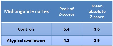
The participants in this study were 9 individuals with atypical swallowing compared to 12 healthy volunteers. Whole-brain resting-state functional images were acquired with a 3.0-T using a 32-channel digital head coil. Echo-planar imaging (EPI) was performed in the axial plane using the following parameters: TR/TE = 2400/30 ms, matrix = 64 × 64, FOV = 192 mm, slice thickness = 3 mm, flip angle = 90°, 45 slices, 4.23 mn scanning time. We used a model-free, independent component analysis (ICA) approach (www.fsl.fmrib.ox.ac.uk/fsl) to generate group-average spatial maps of resting-state brain activity. Two independent raters visually inspected the two group-level spatial maps in order to identify those that best representing the resting-state sensorimotor network (RSSN) based on well-known anatomic network. We performed an additional analysis to assess, mean absolut Z-score and peak of Z-scores within the MCC. For doing so, we created a spherical region-of-interest (ROI) with a radius of 10-mm, approximately positioned to the center of the MCC as defined in the Automated Anatomical Labeling (AAL) template2.RESULTS
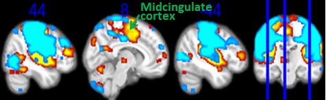
By visually inspecting each group-level ICA component, we were able to identify the components that reflect the RSSN. The figure shows the patterns of low-frequency fluctuations depicted in the group with atypical swallowing (blue-cyan) and control group (red-yellow). Overall, these patterns of activation were similar to that observed in previous studies3 and included regions located along the pre- and post-central gyri, superior frontal gyrus (including supplementary motor area), midcingulate cingulate cortex (MCC), thalamus and insula. However, the control group was found to exhibit more significant activation in the MCC than the group with atypical swallowing (figure). Peak of Z-scores and mean absolute Z-score were higher in the ROI in the controls than in the atypical swallowers (Table).DISCUSSION
Overall, our controls showed greater volume of activation in the MCC compared to the atypical-swallower group, reflecting a high low-frequency synchrony between this area and the remainder regions of the RSSN. This is consistent with results of a previous resting-state functional MRI4 that reported a high functional connectivity between the MCC and the others regions of the cortical swallowing network in healthy individuals. In atypical swallowing, the alterations in lingual functions are likely related to a functional deficit within the swallowing network. Our results suggest that the MCC play a crucial role in the motor control of lingual functions. The MCC was recognized as a site for cognitive control and processing of sensorimotor information 5. Moreover, Whit regard to structural connectivity, the MCC was thought to act as a functional relay in two parallel cerebral loops connecting the sensorimotor and premotor areas with the cerebellum or insula6.CONCLUSION
This work highlights the possibility to investigate functional connectivity of the swallowing network by exploring the synchronicity of intrinsic brain activity within the resting-state sensorimotor network. Our findings suggest that the MCC play a crucial role in the motor control of lingual movements during swallowing. These findings may have important insight for understanding cortical activity in atypical swallowing.Acknowledgements
The authors thank the stall of the Amiens Institute faire Faces for the data acquisitionReferences
1. Locke GR, Talley NJ, Fett SL, Zinsmeister AR, Melton LJ. Prevalence and clinical spectrum of gastroesophageal reflux: a population-based study in Olmsted County, Minnesota. Gastroenterology 1997;112:1448–1456.
2. Tzourio-Mazoyer N, Landeau B, Papathanassiou D, Crivello F, Etard O, Delcroix N, Mazoyer B, Joliot M. Automated anatomical labeling of activations in SPM using a macroscopic anatomical parcellation of the MNI MRI single-subject brain. NeuroImage 2002;15:273–289.
3. De Luca M, Beckmann CF, De Stefano N, Matthews PM, Smith SM. fMRI resting state networks define distinct modes of long-distance interactions in the human brain. NeuroImage 2006;29:1359–1367. doi: 10.1016/j.neuroimage.2005.08.035.
4. Babaei A, Ward BD, Siwiec R, Ahmad S, Kern M, Nencka A, Li S-J, Shaker R. Functional Connectivity of the Cortical Swallowing Network in Humans. NeuroImage 2013;76:33–44. doi: 10.1016/j.neuroimage.2013.01.037.
5. Margulies DS, Kelly AMC, Uddin LQ, Biswal BB, Castellanos FX, Milham MP. Mapping the functional connectivity of anterior cingulate cortex. NeuroImage 2007;37:579–588. doi: 10.1016/j.neuroimage.2007.05.019.
6. Mosier K, Bereznaya I. Parallel cortical networks for volitional control of swallowing in humans. Exp. Brain Res. 2001;140:280–289.
Figures