4540
Working memory decline in patients with amnestic mild cognitive impairment (aMCI) during 2-back tasks1East China Normal University, Shanghai, China, 2Shanghai Ninth People's Hospital, Shanghai, China
Synopsis
This study investigated brain functional abnormalities that are specifically related to working memory in amnestic mild cognitive impairment (aMCI) patients using fMRI in combination with an n-back task. Nineteen aMCI patients (aged, 52-78 years, 13 female) and 19 age-and gender-matched healthy adults participated in this experiment. In the task performance, aMCI group had lower accuracy on the 2-back task. The aMCI patients exhibited less areas of activation during performance of the 2-back vs. 0-back tasks, and more deactivation in the default mode network, compared with healthy group. The aMCI patients exhibited decreased activation in dorsolateral prefrontal cortex and supplementary motor cortex during the 2-back vs. 0-back tasks, compared with healthy group.
Purpose
Mild cognitive impairment include both amnestic and nonamnestic subtypes, and amnestic mild cognitive impairment (aMCI) was highly relevant to Alzheimer’s disease (AD). Patients with aMCI are at risk of developing AD1. Individuals with aMCI show cognitive impairment, especially in memory impairment. Evidence for working memory deficits in aMCI is limited. Investigating working memory in aMCI may help to understand neural changes that occur before there is a profound deficit in behavior. We aimed to identify performance and neural differences between aMCI patients and matched controls on the N-back task.Materials and Methods
Nineteen aMCI patients (aged, 52-78 years, 13 female; MMSE, mean 24.8, SD 1.2) and 19 age-and gender-matched healthy controls (MMSE, mean 29.1, SD 0.7) participated in this experiment. In the current study, n-back task including 3 levels of difficulty (0-back, 1-back, and 2-back). The 0 back require sustained attention, but not working memory, and is typically used as a baseline condition. Functional images during n-back tasks were acquired using an EPI sequence with the following parameters: TR/ TE = 2000/ 30 ms, 172 volumes. Structural scans included a high-resolution three-dimensional T1-weighted magnetization-prepared rapid-acquisition gradient-echo pulse sequence (TR/TE = 2530/ 2.34 ms, 192 slices).One-sample t-tests were used to determine within-group activation for the aMCI and control groups. Two-sample t tests were used to determine between-group differences by SPM12.Results
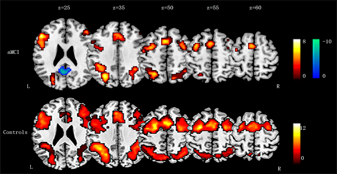
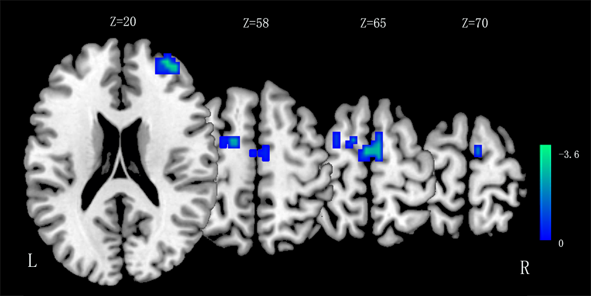
In the task performance, aMCI group showed no significant difference compared to healthy adults during the 0-back or 1-back tasks, but showed poorer accuracy on the 2-back task. The aMCI patients exhibited less areas of activation during performance of the 2-back vs. 0-back tasks, and more deactivation in the default Mode Network (DMN), compared with healthy group, see Figure 1. When compared the two groups directly, aMCI patients exhibited decreased activation in dorsolateral prefrontal cortex ( DLPFC) and supplemental motor area (SMA), see Figure 2.Discussion
In the task performance, aMCI group had low accuracy on the 2-back task. The aMCI group showed less areas of activation during performance of the 2-back minus 0-back tasks compared with healthy group, which suggests that aMCI patients tend to engage less brain areas during 2 back task. In addition, the aMCI patients exhibited more deactivation in the DMN, which suggests that they paid more attention and tried their best to perform the 2-back task. Furthermore, when compared the two groups directly, aMCI patients exhibited decreased activation in DLPFC and SMA, which are important brain regions in the working memory network2. Our results suggests aMCI patients exits potential working memory dysfunction when perform high level n-back task.Acknowledgements
This research was supported by grants from the National Natural Science Foundation of China (NO. 81571658 to X. X. Du)References
1. Schmidtke, K, & Hermeneit, S. High rate of conversion to Alzheimer’s disease in a cohort of amnestic MCI patients. International Psychogeriatrics, 2008;20, 96–108.2. Owen AM, McMillan KM, Laird AR, Bullmore E. N-back working memory paradigm: a meta-analysis of normative functional neuroimaging studies. Hum Brain Mapp 2005; 25:46–59.Figures