4526
Hand-related Auditory and Visual Stimulation on BOLD fMRI and EEG in Children with Autism Spectrum Disorder1Chinese PLA General Hospital, Beijing, China
Synopsis
By using Blood oxygen level dependent (BOLD) magnetic resonance imaging (MRI) method with hand-related pictures and sound stimulating on Autism Spectrum Disorder (ASD) children, the difference was found in the border of temporal lobe and occipital lobe in ASD and NC contrast. And these findings may help guide the precise intervention or treatment in ASD.
Abstract
Purpose: The present study aimed to expand current knowledge regarding the early diagnosis of ASD and potentially discover the pathological mechanisms underlying psychiatric developmental disorders.
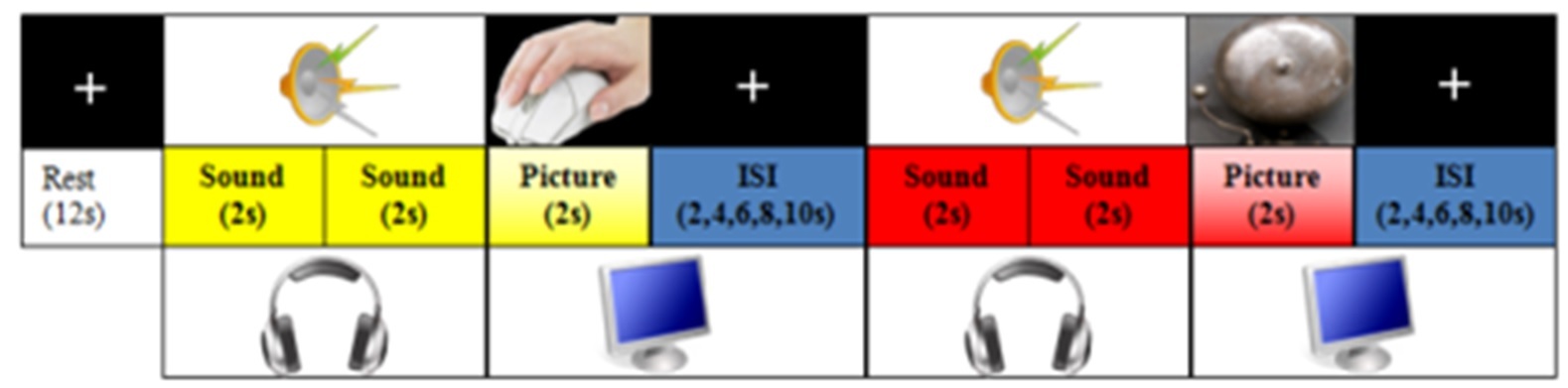
Methods: An institutional review board approved the present retrospective case-control study, and all participants and their guardians provided informed consent. BOLD imaging was utilized in conjunction with a hand-related auditory and visual stimulation experiment to assess 20 ASD participants and 20 age-matched NC participants. Diagnoses of ASD were based on Diagnostic and Statistical Manual of Mental Disorders, Fourth Edition (DSM-IV), Global Assessment of Functioning (GAF) scores (mean score: 8.00 ± 0.50). For the NC group, 20 typically developing (TD) children with no previous history of neurological or psychiatric problems were recruited as controls. All participants had normal or corrected-to-normal vision, and no participants reported the use of psychotropic medications. The exclusion criteria were metal implants, psychiatric or neurological disorders, structural brain abnormalities, and known genetic conditions. MRI scans were read as normal by experienced neuroradiologists, and none of the children exhibited any signs of prematurity-associated brain lesions on MRI at a term-equivalent age, as assessed by case report forms. The study protocol was approved by the Medical Ethics Committee of the Chinese PLA General Hospital and was fully disclosed to all participants and their guardians; written informed consent was obtained from each participant’s guardian according to the Declaration of Helsinki. T2-weighted images (T2WI), three-dimensional fast spoiled gradient echo (3D-FSPGR) images, and BOLD images were obtained to statistically analyze the spatial distribution. The BOLD scan parameters were as follows: TR = 2000 ms, TE = 30 ms, NEX = 1, acquisition matrix = 64 × 64, FOV = 240 × 240 mm, thickness = 3.8 mm, and 36 slices. All participants underwent MRI scans on a 3.0T MRI system (Discovery MR750 system), and the event-related designs were composed of four runs per participant, with each run lasting 252 s. Each run included 20 trials: One trial lasted 12 s and included 2 s of sound, 2 s of sound, 2 s of a picture, and a 6-s interstimulus interval; the dummy scan lasted 12 s (Fig. 1). The stimulation programs were applied using E-Prime2.0 Standard and the China Shenzhen Sinorad system. All hand-related and non-hand-related pictures and sounds conformed to Hemera Photo Objects Database requirements in terms of frequency, power, and resolution. Matlab and the SPM8 plugin were used to assess BOLD data.
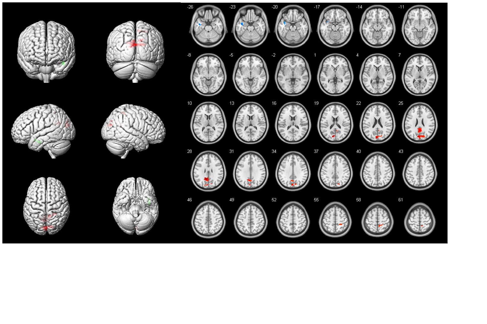
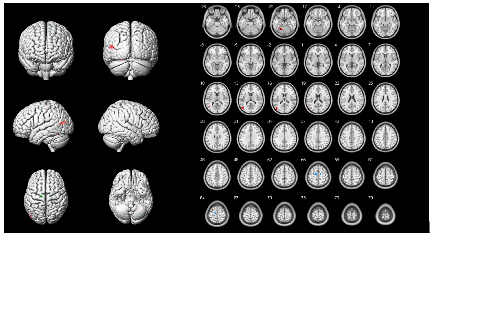
Results: The present study found a difference in contrast on the border of the temporal lobe and occipital lobe between the ASD and NC groups: (1) auditory stimulation fMRI BOLD results (n = 3; Figs. 2a, b, d, e, g, and h; P < 0.05, family wise error [FEW]; Figs. 2c, f, and i; P < 0.01), (2) hand-related visual stimulation BOLD fMRI results (Fig. 3, L: 3D, P < 0.05), and (3) hand-related auditory stimulation BOLD fMRI results (Fig. 4, L: 3D, P < 0.05).
Conclusions: The present study aimed to localize brain abnormalities in preschool-age children with ASD, uncover the molecular mechanisms underlying the stereotyped behaviors observed in ASD children, and illustrate the importance of fMRI assessments in the treatment of ASD. Future research from our group will use novel stimuli for ASD auditory comprehension assessments and apply BOLD fMRI in a larger sample.
Acknowledgements
Financial support was received from the National Natural Science Foundation of China (31100714), Beijing Natural Science Foundation (7162183), and Beijing Nova Program (xxjh2015105; 2011112).References
[1] Javad F, Warren JD, Micallef C, et al. Auditory tracts identified with combined fMRI and diffusion tractography. Neuroimage, 2014; 84: 562–574.
[2] Medeiros K & Winsler A. Parent-child gesture use during problem solving in autistic spectrum disorder. Journal of Autism and Developmental Disorders, 2014; 44(8): 1946–1958.
[3] O’Nions E, Sebastian CL, McCrory E, et al. Neural bases of theory of mind in children with autism spectrum disorders and children with conduct problems and callous-unemotional traits. Developmental Science, 2014, 17(5): 786–796.
Figures