4523
Silent Simultaneous EEG-fMRI using Looping-Star.1TUM (Technical University of Munich), Munich, Germany, 2GE Healthcare, Munich, Germany, 3Max Planck Institute of Psychiatry, Munich, Germany
Synopsis
This work presents a novel technique for silent simultaneous EEG-fMRI, combined with a slow gradient switching fMRI method called Looping-Star. In comparison to the conventional setup with Gradient-Echo EPI, the silent technique improves the EEG quality while maintains fMRI quality characteristics. The inaudible scanning offers higher sensitivity to auditory stimuli, resulting in enhanced evoked response potentials (ERPs).
INTRODUCTION
The high acoustic noise caused by the rapid gradient switching from the echo planar imaging (EPI) sequences is especially unsuitable for simultaneous EEG-fMRI1-3. This work presents a novel technique with unique silent characteristics, which combines simultaneous EEG with a slow gradient switching fMRI method called Looping-Star4-6. The new technique is evaluated in terms of data quality (in phantom and in-vivo experiments) and sensitivity to functional BOLD responses during an auditory stimulation paradigm in comparison with standard Gradient Echo EPI.METHODS
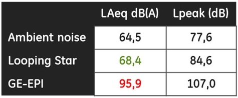
In-bore acoustic noise measurements were performed with a Bruel and Kjaer Type-4189 calibrated microphone placed inside the head coil during 30s for each setup and ambient noise.
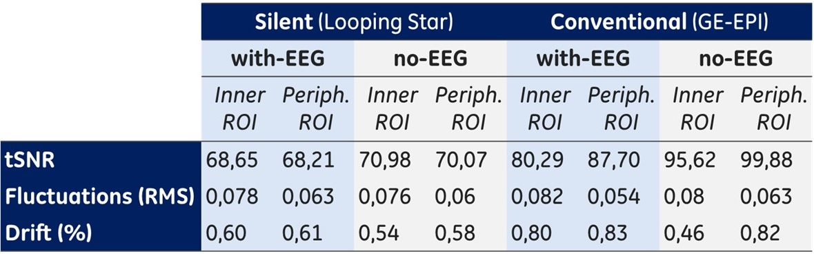
To evaluate the quality of the data acquired with the silent technique, a spherical water phantom with a 32-channels EEG BrainCap (Brainproducts, Munich, Germany), was imaged in a GE 3T MR750w (GE Healthcare, Waukesha, WI, USA), with two scan protocols: T2* Looping-Star (FA=1°, BW=±31.25 kHz, FOV=19.2 cm, 3 mm isovoxel, 64x64, TE 26.88ms, TR 2.24s, 100 volumes), and GE-EPI (FA=80°, BW=±31.25 kHz, FOV=19.2cm, 3mm isovoxel, 64x64, TE=26.88ms, TR=2.24s, 100 volumes) for comparison. Induced gradient artifacts (GA) were analyzed off-line, comparing the artifact pattern, amplitude and spectral components for each sequence using Brain Vision Analyzer 2.0. Additionally, temporal stability measurements7 were calculated over two proportional ROIs8: inner region and peripheral region of the phantom, with and without EEG cap.
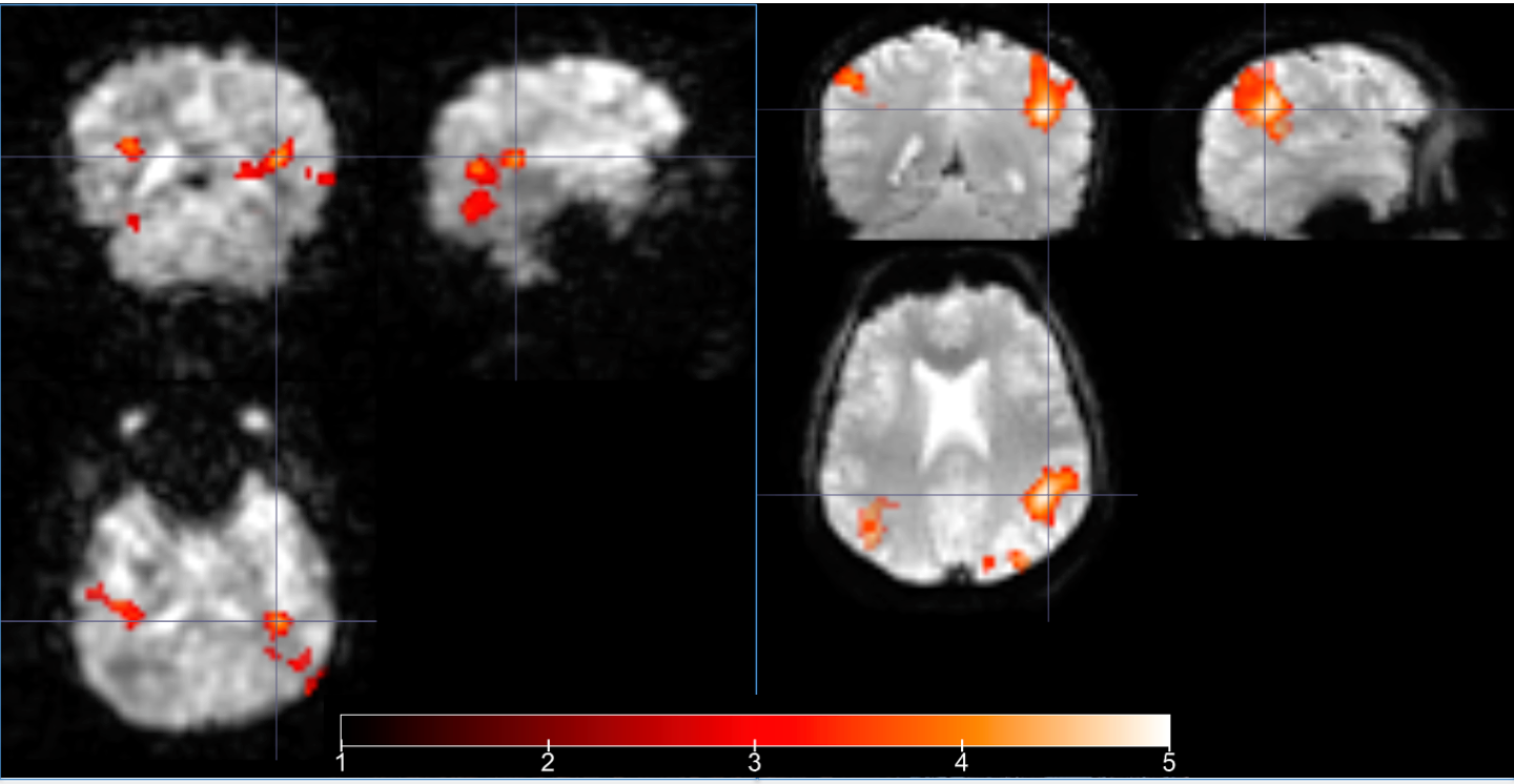
To address the sensitivity of the silent technique to functional responses, one healthy volunteer was scanned during an auditory oddball experiment (1.5KHz odd tone (20% probability), 1KHz frequent tone, duration=0.5s, ISI=2s, random jitter of 0, ±100 or ±200 ms). The experiment was performed during 10 minutes for three scenarios: a) with T2* Looping Star, b) with GE-EPI and c) outside the bore.
BOLD activation spatial maps were computed with a GLM analysis, including two regressors (standard and odd tones onsets) and the canonical HRF as convolution. When comparing all rare tone against standard tones, significance was set to p<0.05 and Z>2.3, cluster-wise FWE corrected.
RESULTS
A 40,2% reduction in sound intensity was obtained with Looping Star compared to GE-EPI (Table 1).
The silent method improves the overall EEG data quality, since Looping Star induced GA showed a smoother pattern with significantly reduced amplitude in all electrodes, a factor of 2 lower than GE EPI, (Figure 1) and a uniform noise spectrum.
Both setups were similar in terms of the fMRI data quality (Table 2). The introduction of the EEG system causes a minor loss of tSNR and increased temporal drifts, with slightly higher effects on the GE-EPI images, markedly in the peripheral region of the phantom.
Figure 2 illustrates the results of the auditory ERPs obtained during the oddball paradigm. The waveforms show a clear enhanced response in the silent setup. The P100 and N200 amplitudes are higher than in the conventional setup and close to the out-bore acquisition.
fMRI activations resulting from the differential contrast (odd>frequent tones) were found for both setups (Figure 3), predominant in brain regions involved in selective attention (fronto-parietal, temporal and superior motor area).
DISCUSSION
In this work, we report our preliminary experience with a silent method for simultaneous EEG-fMRI using Looping Star. In summary, we demonstrated that: (i) the silent setup improves the EEG quality with significantly reduced gradient artifacts, while maintaining overall similar fMRI quality characteristics; (ii) ERP signals are higher for the silent relative to the conventional setup, suggesting a higher sensitivity to auditory stimuli and potentially higher SNR, which may be extended to the single trial level; (iii) the novel technique offers sufficient BOLD sensitivity to detect event related responses, while providing 3D brain coverage. Further experiments with an extended sample and non-auditory tasks should be conducted to verify these results.Acknowledgements
No acknowledgement found.References
[1] Le et al., Magn Reson Med (2001)
[2] Amaro et al., J. Magn. Reson. Imaging (2002)
[3] Moelker et al., MAGMA (2003)
[4] Solana Sanchez et al., ISMRM (2016)
[5] Solana Sanchez et al., ISMRM (2017)
[6] Wiesinger et al., ISMRM (2017)
[7] Friedman and Glover, J Magn Reson Imaging (2006)
[8] Mullinger et al., Int J Psychophysiol (2007)
Figures