4519
Improving Sensitivity of Task-fMRI Signals by Use of Time Delayed Systemic Regressors: A Comparison of Probe Regressors from Peripheral NIRS Recordings and BOLD-fMRI1Medical Engineering, Acibadem Mehmet Ali Aydinlar University, Istanbul, Turkey, 2College of Engineering, Purdue University, West LaFayette, IN, United States, 3Department of Radiology, University of Calgary, Calgary, Alberta, Canada, 4Department of Psychiatry, Harvard University Medical School, Belmont, MA, United States, 5Department of Psychiatry, Harvard Medical School, Belmont, MA, United States
Synopsis
A fundamental problem with fMRI measurements is the strong presence of low frequency systemic physiological noise (<0.15 Hz), which significantly corrupts detection power for hemodynamic variations caused by task induced neuronal activation. In this study, we propose a novel noise removal strategy for task-fMRI studies by taking into consideration a relatively new established property of systemic low frequency oscillations (sLFOs): their dynamic propagation within cerebral vasculature causing voxel-specific arrival delays. We compare the performance of dynamic noise modelling regressors obtained from i) BOLD data and ii) a fingertip HBO signal of non-neuronal origin concurrently recorded with near infrared spectroscopy (NIRS).
Introduction
Functional magnetic resonance imaging (fMRI) is a commonly used brain imaging modality with a wide range of promising clinical applications. A fundamental problem with fMRI measurements is the strong presence of low frequency systemic physiological noise ( < 0.15 Hz), which significantly corrupts detection power for hemodynamic variations caused by task-induced neuronal activation [1]. Recent studies demonstrated that systemic low frequency oscillations (sLFOs) in resting state BOLD data account for up to 30% of the variance in gray matter [2,3] which also strongly decreases the contrast-to-noise ratio (CNR) in task-fMRI studies by causing false negatives and false positives in brain activation maps. Previous work from our group identified important characteristics of the sLFOs in resting state BOLD-fMRI data. sLFOs were shown to be intrinsic blood borne signals that propagated dynamically both in peripheral tissues with site dependent time delays [4] and throughout the brain with cerebral blood circulation [5-7]. In the present study, we evaluate the impact of sLFOs on task-activation studies and investigate the extent to which sLFOs affect task-fMRI data in both activated and non-activated brain regions. We propose a novel noise removal method for task-fMRI data by taking into account voxel-specific arrival time of sLFOs. We model physiological noise in BOLD signals collected during a serial subtraction task with systemic waveforms derived from i) BOLD data itself and ii) a fingertip HBO signal concurrently recorded with near infrared spectroscopy (NIRS). We utilize each noise modeling regressor in a general linear model (GLM) by applying an optimal time delay computed for each voxel prior to regression. We then compare the performance of each regression method in improving a) functional contrast to noise ratio (CNR), b) sensitivity and c) specificity of the brain activation maps at the single subject and group level. Our results suggest that time delay processing of the global signal derived from voxels excluding an activation mask obtained after standard fMRI preprocessing steps explains variance to a better extent, and results in improved activity localization with higher sensitivity and specificity.Methods
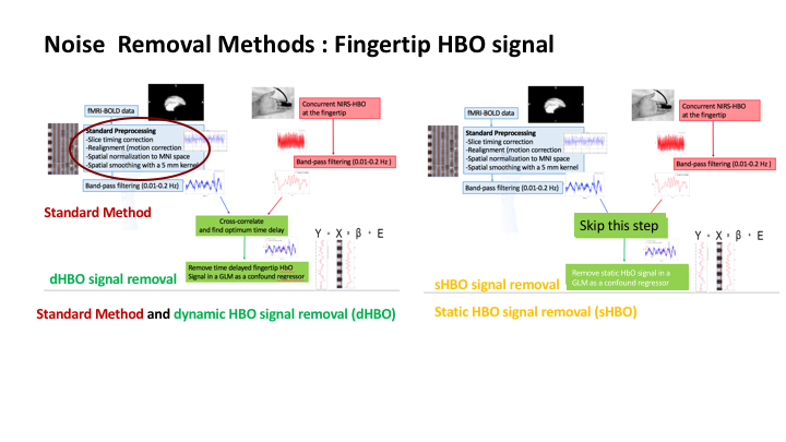
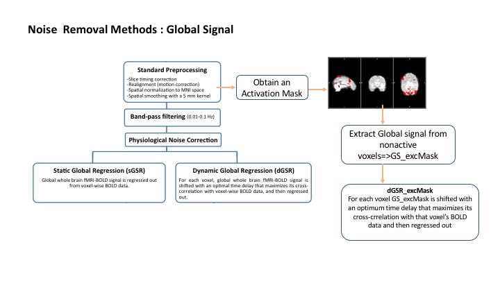
A concurrent resting state NIRS-fMRI scan was followed by a 4 minute serial subtraction experiment separated by 2 minutes of rest. Functional data was preprocessed using FEAT, (http://fsl.fmrib.ox.ac.uk). A temporal bandpass filter was applied to retain frequencies in the range of 0.01-0.2 Hz. NIRS data was collected with a probe placed on the right fingertip (Imagent, ISS, Inc., Champaign, IL). Noise Removal Procedures: Standard Method: A multiple linear regression analysis was performed with the standard preprocessing steps depicted in Fig.1. Static HbO Regression(sHBO): The fingertip NIRS-HBO signal was used as a confound regressor in a general linear model (GLM) analysis (Fig.1). Dynamic HbO Regression(dHBO): For each voxel BOLD time series, the noise modeling regressor was obtained by shifting the fingertip NIRS HBO signal with an ‘optimal’ time delay that maximizes its cross-correlation with that voxel’s BOLD fMRI time series (Fig.1). The voxel-specific optimal time delay was determined using the Regressor Interpolation at Progressive Time Delays (RIPTiDe) procedure with in-house built, custom software [8].Static Global Signal Regression(sGSR): The global signal is used as a regressor for removing systemic effects in a GLM analysis. Dynamic global signal regression(dGSR): A voxel-specific optimally delayed version of global signal is used as a regressor in the GLM utilizing RIPTiDe procedure [3,8]. Optimized dynamic global signal regression (dGSR_excMask): Standard preprocessing steps are applied to fMRI data and a brain activation mask is obtained. Global signal is calculated from voxels outside this initial brain activation mask and denoted as 'GS_excMask'. The same procedure for dGSR is applied to GS_excMask [8].
Results and Discussion
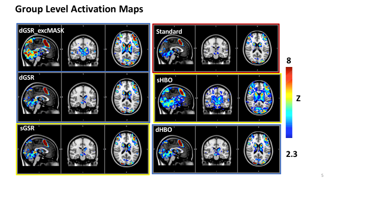
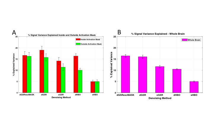
Fig. 3 demonstrates the performance of each method in explaining variance A) inside and outside the activation masks, and B) from the whole brain. dGSR approaches explain variance to a greater extent (up to %15) than the other methods with similar performances for voxels inside and outside the activation mask. However; dGSR_excMask results in significantly higher CNR for voxels deemed significant after noise removal (Fig. 4A) while presenting significantly higher specificity than dGSR in depicting specificity to detecting task-induced hemodynamic variations (Fig. 4B). These results are also supported with z-scores of brain activation obtained at the group level (Fig.5). Denoising fMRI data with voxel-specific optimally aligned global signal obtained from non-active brain voxels resulted in a better localized activation area with a higher CNR within the corresponding positive activation map when compared to analysis with other approaches.Conclusion
We suggest that for task fMRI studies, a preprocessing pipeline utilizing time delay processing of the global signal derived from voxels excluding an activation mask obtained after standard fMRI preprocessing steps results in more variance removal and improved activation localization with higher sensitivity and specificity.Acknowledgements
This work was supported by the National Institutes of Health, Grants K25 DA031769(YT), and R21 DA032746 (BdeBF). SBE was supported by the BIDEB 2219 program of the Scientific and Technological Research Council of Turkey (TÜBITAK).References
[1] M. Bianciardi, M.Fukunaga, P. van Gelderen, S.G. Horovitz, J.A. de Zwart, K. Shmueli, et al., “Sources of functional magnetic resonance imaging signal fluctuations in the human brain at rest: a 7 T study, ”Magn. Reson. Imaging, ” 27 , pp.1019–1029, 2009. [2] B.D Frederick, L.D. Nickerson and Y. Tong, “Physiological denoising of BOLD fMRI data using Regressor Interpolation at Progressive Time Delays (RIPTiDe) processing of concurrent fMRI and near-infrared spectroscopy (NIRS) ,” NeuroImage. 60(3):1913–1923, 2012. [3] S.B. Erdoğan, Y. Tong, L.M. Hocke, K.P. Lindsey and B.D. Frederick, “Correcting resting state fMRI-BOLD signals for blood arrival time enhances functional connectivity analysis,” Front. Hum. Neurosci., 28 June 2016 http://dx.doi.org/10.3389/fnhum.2016.00311. [4] Y. Tong, L.M. Hocke, S.C. Licata and B. Frederick, “Low-frequency oscillations measured in the periphery with near-infrared spectroscopy are strongly correlated with blood oxygen level dependent functional magnetic resonance imaging signals,” J Biomed Opt 17:106004, 2012. [5] L.M. Hocke, Y. Tong, K.P. Lindsey and B.D Frederick, “ Comparison of peripheral NIRS LFOs to other denoising methods in resting state functional fMRI with ultra-high temporal resolution,” Magn. Reson. Med. 2016 Feb 7 doi: 10.1002/mrm.26038. [6]Y. Tong and B.D. Frederick, “ Time lag dependent multimodal processing of concurrent fMRI and near-infrared spectroscopy (NIRS) data suggests a global circulatory origin for low-frequency oscillation signals in human brain,” NeuroImage 53(2):553–564, 2010. [7] Y. Tong, K.P. Lindsey and B.D. Frederick, B.D., “Partitioning of physiological noise signals in the brain with concurrent near-infrared spectroscopy and fMRI,” J. Cereb. Blood Flow Metab. 31:2352– 2362, 2011. Y. Tong and B.D. Frederick, “ Tracking cerebral blood flow in BOLD fMRI using recursively generated regressors,” Hum. Brain Mapp. 35, 5471–5485, 2014. [8]https://github.com/bbfrederick/rapidtide
Figures
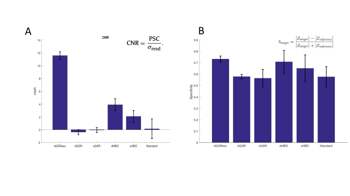
Improvements in A) CNR and B) Specificity for each noise removal method.