4505
A study on Compressed SENSE(CSENSE) in prostate T2map at 3T.seiichiro noda1, nobuyuki toyonari1, yukari horino1, kazuhiro katahira1, masami yoneyama2, and yasutomo katsumata3
1kumamoto chuo hospital, kumamoto, Japan, 2philips japan, tokyo, Japan, 3pilips japan, tokyo, Japan
Synopsis
We study on Compressed SENSE in prostate T2map at 3T The imaging sequence used spin echo type multi echo. In case of SENSE, increase Factor resulted in an artifact peculiar to parallel imaging and an error occurred in T2 value measurement of the prostate. However, in the case of CSENSE, artifacts were reduced and T2 value measurement was also possible. Prostate T2map can reduce imaging time using CSENSE.
Synopsis
We study on Compressed SENSE in prostate T2map at 3T The imaging sequence used spin echo type multi echo. In case of SENSE, increase Factor resulted in an artifact peculiar to parallel imaging and an error occurred in T2 value measurement of the prostate. However, in the case of CSENSE, artifacts were reduced and T2 value measurement was also possible. Prostate T2map can reduce imaging time using CSENSE.Purpose
Diffusion weighted images are widely used in the diagnosis of prostate cancer. It is reported that ADC is useful for diagnosis of malignancy because it is excellent in quantitativeness. However, because the diffusion weighted image is EPI, distortion is caused by the influence of rectal gas, which may interfere with ADC measurement. This time, we focused on T2map using spin echo type multi echo. It is suggested that possibilities for diagnosis of prostate cancer may be useful also in past papers and the like. However, there is concern that the imaging time is long. SENSE is common as a method to reduce imaging time. Increasing SENSE factor and imaging ProstateT 2 map causes error in T 2 value measurement due to SENSE artifact. (Fig.1) On the other hand, CSENSE can undersample collected data. It is a technology that can shorten the imaging time. We examined whether CSENSE can be used for prostate T2map.Methods
PHILIPS 3.0T Ingenia R5.4 was used as the equipment to be used. Experiments T2map was imaged for 5 conscious healthy volunteers. We picked up CSENSE reduction factor 3 (scan time 4 min 20 sec) and 6 (scan time 2 min 15 sec). From the obtained T2map, the T2 values of reduction factors 3 and 6 of the prostatic gland and the internal gland were measured and compared. T-test. Imaging was performed on T2map reduction factor 6 (scan time 2 min 15 sec) for prostate cancer patients who got consent. Parameter optimization: FOV 200 mm, RFOV 100, Oversampring 90 mm, Matrix 112, Recon 224, scan% 70, NSA 1, 2 D multi slice, TR 2500 ms, multi echo (TE 20 ms to 160 ms) 20 ms interval 8 echo is acquired. Thickness 4mm, 16 slice T2map was created.Results
Artifacts are unlikely to occur even if CSENSE reduction factor 3 is changed to 6. T2 value measurement is also possible. For reduction factor 3, the external gland T2 value is 214.86 ± 52.1 ms and the internal gland is 99.6 ± 18.4 ms. In the case of reduction factor 6, the external glands were 222.66 ± 56 ms and 99.5 ± 16.9 ms. (Fig.2)Discussion
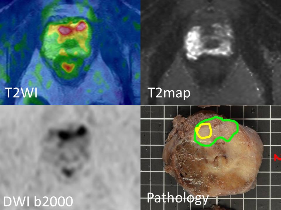
The t-test of the external gland T2 value was 0.0127> 0.01 p, and there was some variation. It is thought that it is an influence of respiration at the time of imaging and movement of the intestinal tract. In the case of the internal glands, there is no significant difference and it is considered that there is little influence on this imaging. In order to incorporate T2map into prostate routine examination, extension of imaging time has concerns such as body movement. This time, it was possible to scan for about 2 minutes. We did not use the Grace method to consider the influence of the magnetic susceptibility, but in the future comparison and examination is necessary. Imaging was also performed on reduction factor 6 (scan time 2 min 15 sec) for the patient who got consent. View images.(Fig,3)Conclusion
Prostate T2map can reduce imaging time by using CSENSE. There was T2 value measurement was also possible.Acknowledgements
No acknowledgement found.References
1、Accelerated T2 Mapping for Characterization of Prostate Cancer Wei Liu, D.Sc.1,2,‡, Baris Turkbey, M.D.2,‡, Julien Sénégas, Ph.D.3, Stefanie Remmele, Ph.D.3, Sheng Xu, Ph.D.1, Jochen Kruecker, Ph.D.1, Marcelino Bernardo, B.S.4, Bradford J. Wood, M.D.5, Peter A. Pinto, M.D.5, and Peter L. Choyke, M.D.2 Magn Reson Med . 2011 May ; 65(5): 1400–1406. doi:10.1002/mrm.22874. 2、Prostate Cancer Discrimination in the Peripheral Zone With a Reduced Field-of-View T2-mapping MRI Sequence Fernando I. Yamauchi, MD1,2, Tobias Penzkofer, MD1,3, Andriy Fedorov, PhD1, Fiona M. Fennessy, MD, PhD4, Renxin Chu, PhD1, Stephan E. Maier, MD, PhD1,5, Clare M.C. Tempany, MD1, Robert V. Mulkern, PhD1,6, and Lawrence P. Panych, PhD Magn Reson Imaging. 2015 June ; 33(5): 525–530. doi:10.1016/j.mri.2015.02.006. 3、Characterization of prostate cancer using T2 mapping at 3T : A multi scanner study A. Hoang Dinh 4、Development of fast imaging techniques in MRI − From the principle to the recent development − Yoshio MACHIDA and Issei MORI Health Sciences, Tohoku University Graduate School of Medicine 2-1 Seiryo-machi, Aoba-ku, Sendai, 980-8575, Japan (Received on October 13, 2012)Figures
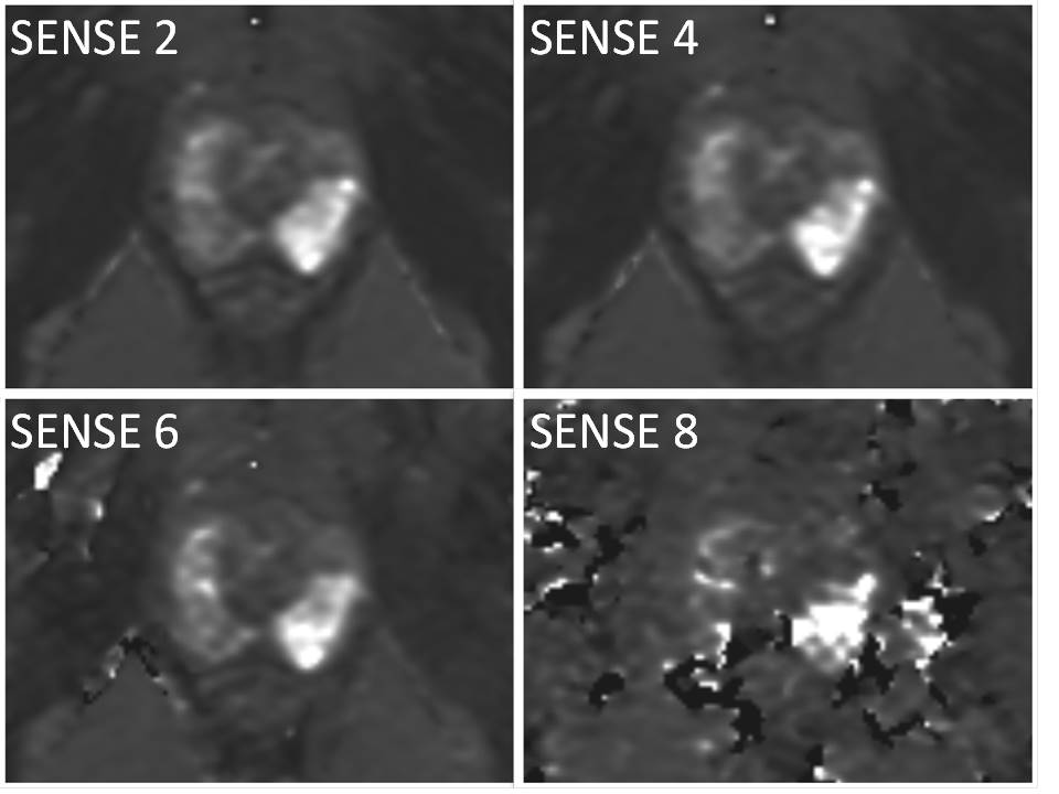
In case
of SENSE, increase Factor resulted in an artifact
peculiar to parallel imaging and an error occurred in T2 value measurement of
the prostate.
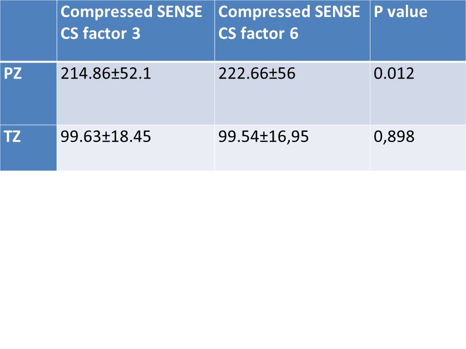
We picked up CSENSE reduction factor
3 (scan time 4 min 20 sec) and 6 (scan time 2 min 15 sec). From the obtained
T2map, the T2 values of reduction factors 3 and 6 of the prostatic gland and
the internal gland were measured and compared. T-test.
P Value tests the difference between the T2
values measured on the CS factor
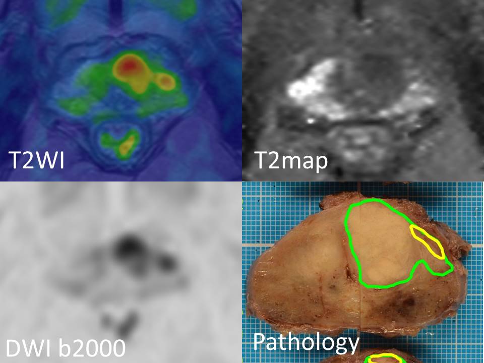
In the ventral region
of the prostate, low signal area at T2WI, abnormally high signal on diffusion
weighted image, findings of prostate cancer are recognized.
Moderately diff. Adenoca., Gleason score 3 + 4
PSA = 10.32
Low signal on T2WI on the ventral side of prostate apex and high signal on
diffusion weighted image are recognized.
Prostate cancer at the same site is suspected.
Moderately diff. Adenoca., Gleason score 3 + 4, Stage B 2