4467
Depiction of Gas Flow Patterns in Pulmonary Dynamic Hyperpolarized Gas MRI1State Key Laboratory of Magnetic Resonance and Atomic and Molecular Physics, National Center for Magnetic Resonance in Wuhan, Wuhan Institute of Physics and Mathematics, Chinese Academy of Sciences, Wuhan, China, 2University of Chinese Academy of Sciences, Beijing, China
Synopsis
Dynamic hyperpolarized gas MRI of lung is able to visualize the dynamic changes of pulmonary morphology during the ventilation process, which provides the important information about lung physiology and pathophysiology. However, there is a lack of an effective method to depict the gas flow patterns in the whole lung. In this work, we propose a motion field calculation method based on the pulmonary gas flow properties, which can acquire the velocity and direction of gas flow. The performance of the proposed method is investigated experimentally.
Introduction
Hyperpolarized (HP) gases (such as 3He and 129Xe) MRI have shown great potential in the early diagnosis of lung diseases because of the capability to evaluate both pulmonary structure and function.1,2 In order to visualize the dynamic changes of pulmonary structure during the ventilation process, dynamic HP gas MRI of the lung was developed, which enables to provide the information about the lung physiology and pathophysiology.3 Gas flow in human airways has also been measured successfully.4 However, there is not an effective method to depict the gas flow patterns in the whole lung, which may give an insight into the lung, such as the airways, alveolar and function of gas exchange.
The gas flow in the lung can be considered as a non-rigid deformation and restricted motion that extent of restriction changes in the different position of lung. Therefore, we propose a motion field calculation method based on these pulmonary gas flow properties, which acquires the velocity and direction of gas flow from the pulmonary dynamic HP gas MR images.
Methods
The motion field calculation method can be divided into four steps. First, dynamic images of lung are acquired through dynamic HP gas MRI. Second, each frame was prefiltered using an anisotropic filter to reduce the noise, while at the same time preserving the details. This filtering operation constitutes a trade-off between improvement in signal-to-noise ratio and preservation of image details. Third, the motion field of gas flow are calculated by considering the image sequence as the brightness function. The motion filed of gas-flow locally satisfies the brightness conservation law (dE/dt = 0). Moreover, the restriction extent of gas flow changes in the different position of lung, leading to a variation of brightness. This suggests that the magnitude of velocity can be described as a function of brightness. Based on the above, one can acquire the motion field by solving the following optimization problem,
$$\min_{v_{x}, v_{y}}\parallel\frac{\partial E}{\partial t}+\frac{\partial E}{\partial x}\cdot v_{x}+\frac{\partial E}{\partial y}\cdot v_{y}\parallel_2^2+\lambda_{1}\parallel v- f(E)\parallel_2^2, v=(v_{x}^2+v_{y}^2)^\frac{1}{2}$$
where the vx and vy are the velocity components of the motion filed in the x and y directions, v is the magnitude of velocity, λ1 is the regularization parameter to balance the brightness conservation law and the gas flow characteristics in the lung, f is the function to describe the correlation between the brightness and magnitude of velocity. After the solution of the proposed objective function, the motion filed is acquired. In the last step, the motion filed is overlapped with each dynamic HP gas MR image. In this way, the velocity and direction of HP gas flow in whole lung is acquired.
To investigate the feasibility and robustness of the proposed method in vivo, dynamic HP 129Xe MRI experiments were performed on a 1.5 Tesla whole-body MRI scanner (Avanto, Siemens Medical Solutions). Six healthy volunteers were enrolled. A homebuilt transmit-receive vest radio frequency coil was used in the experiments. The imaging parameters were as follows: the matrix size = 128×128, frame number = 15, TR/TE = 10.5/5 ms, field of view = 384×384 mm2, 2D FLASH sequence, acceleration factor = 3. After the acquisition of HP gas MR data, images were reconstructed using an algorithm with low-rank and sparsity constraints. Then the motion field calculation method was performed on the images.
Results
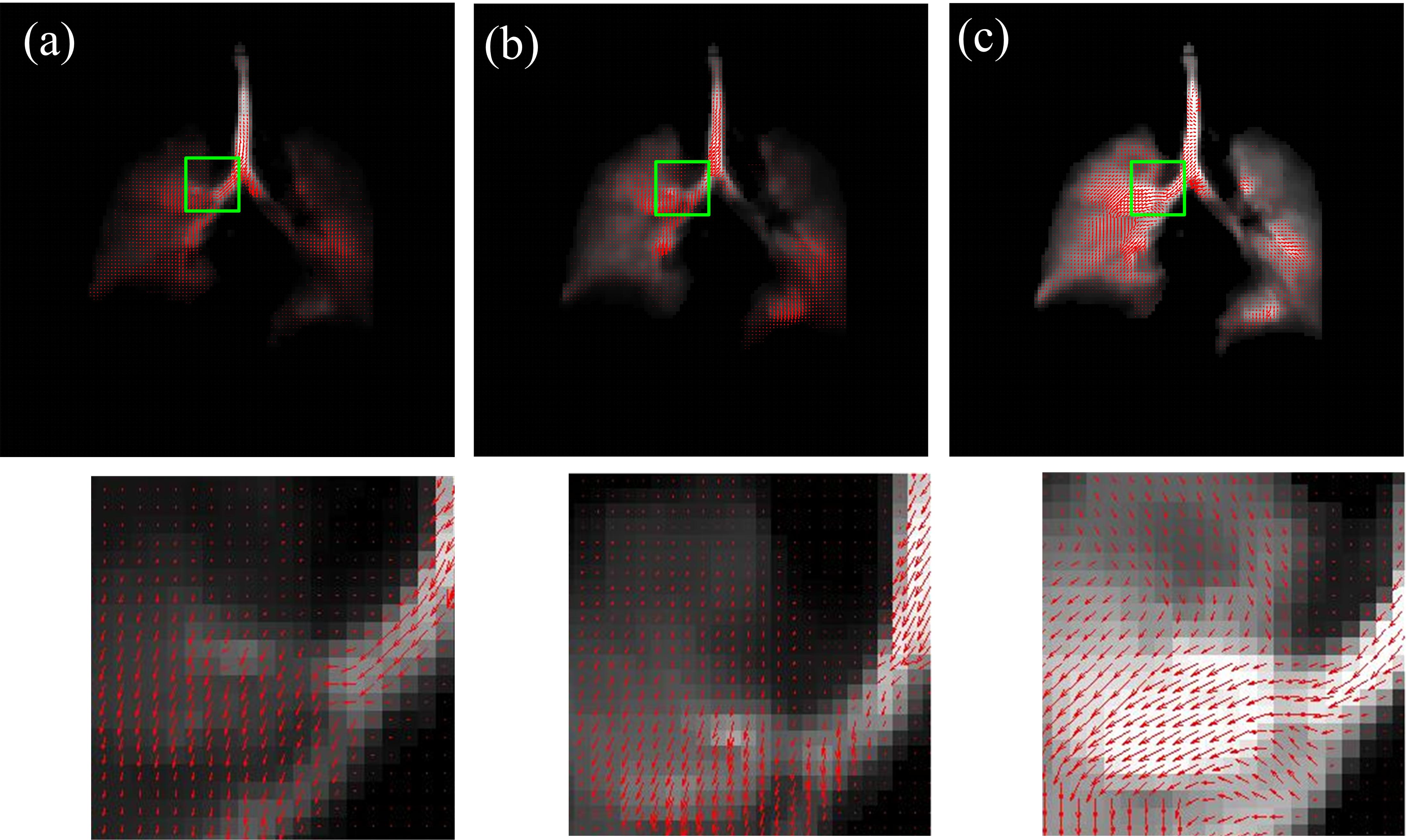
The motion filed calculation method was successfully implemented in the experiment. Figure 1 demonstrates gas flow velocity and direction in 1st, 5th, 9th and 13th frames of one volunteer. In order to show the images more clearly, regions of interests were zoomed in (indicated by The green rectangles). It can be seen that the gas flow dynamics in the whole lung were clearly visualized in this volunteer over the 6.67 s duration of the experiment.Discussion
Some features are shown in the results. There are continuities in the motion field maps of adjacent frames. In addition, the velocity of gas flow in the trachea and main bronchi is higher than that in other positions of lung. These features are consistent with the gas flow properties in the lung, demonstrating the feasibility of the proposed method. Moreover, similar features are also observed in results of other five volunteers, demonstrating the robustness of the proposed method.Conclusion
This work has demonstrated that the proposed method can successfully be applied to calculate the motion filed of gas flow. This provides an efficient way to dynamically depict the gas flow patterns in the whole lung, which is helpful to analyze the lung physiology and pathophysiology.Acknowledgements
This work was supported by the National Natural Science Foundation of China (81227902, 61471355, 81625011, 81771917). XZ acknowledges the support by the National Program for Support of Eminent Professionals (National Program for Support of Top-notch Young Professionals).References
1.Albert MS, Cates GD, Driehuys B, Happer W, Saam B, Springer CS, Wishnia A. Biological magnetic-resonance-imaging using laser polarized Xe-129. Nature 1994; 370(6486):199-201.
2.Li H, Zhang Z, Zhao X, Sun X, Ye C, Zhou X. Quantitative Evaluation of Radiation-induced lung injury with hyperpolarized xenon magnetic resonance. Magn Reson Med 2016;76(2):408-416.
3.Viallon M, Berthezene Y, Callot V, et al. Dynamic imaging of hyperpolarized 3He distribution in rat lungs using interleaved-spiral scans. NMR Biomed 2000;13(4):207-213.
4.Collier GJ, Wild JM. In vivo measurement of gas flow in human airways with hyperpolarized gas MRI and compressed sensing. Magn Reson Med 2015;73(6): 2255-2261.
Figures